
Abstract
Advanced abdominal pregnancy is a rare but life-threatening condition associated with substantial maternal and fetal morbidity and mortality, particularly when diagnosis is delayed beyond the first trimester. Owing to its rarity, there is still no consensus regarding its optimal diagnostic approach or management. We present a case of advanced abdominal pregnancy in a woman who first sought medical care at 24 weeks’ gestation. She reported five months of amenorrhea accompanied by progressively worsening abdominal pain, constipation, and dyspareunia. Ultrasonography demonstrated a gestational sac outside the uterine cavity. Magnetic resonance imaging was subsequently performed to define the placental location and its relationship to adjacent structures and to confirm the diagnosis of advanced abdominal pregnancy. Because the patient had persistent severe abdominal pain and clinical findings concerning for an acute abdomen, immediate surgical management was undertaken after counseling and preoperative planning. Laparotomy was performed through a midline abdominal incision. Intraoperatively, the omentum, uterus, adnexa, and bladder were densely adherent to the gestational sac, creating the appearance of a frozen pelvis. The placenta invaded the peritoneum overlying the sigmoid colon and left iliac region, although no major vascular involvement was identified. Organ-sparing surgery was successfully performed, and the placenta was carefully dissected and removed without complications. The postoperative course was uneventful, and the patient was discharged in good condition. Advanced abdominal pregnancy should be considered in the differential diagnosis of pregnant patients presenting with abdominal pain and atypical imaging findings. In symptomatic patients with an acute abdomen, immediate surgery may be preferred over expectant management. Although placental management remains controversial, detailed preoperative imaging is crucial for surgical planning. In selected cases without major vascular invasion, placental removal may be feasible with organ preservation.
Keywords: Advanced abdominal pregnancy, ectopic pregnancy, placenta, magnetic resonance imaging, surgical procedures
Introduction
Ectopic pregnancy is defined as the placement of the blastocyst outside the endometrium and is seen in approximately 1-2% of cases(). Abdominal pregnancy (AP), which constitutes 1% of all ectopic pregnancies or 1/10,000-30,000 of all pregnancies, is the placement of the placenta anywhere in the peritoneal cavity(-). When the gestational age is less than the 20th week of pregnancy, AP has a prognosis and treatment options similar to those for other ectopic pregnancies. However, in cases of advanced AP (AAP), where the gestational age is over 20 weeks, the prognosis is worse than that of other ectopic pregnancies. Two important factors that determine prognosis are gestational age at diagnosis and the location of the placenta. Because AAPs are rare, their treatment is controversial(). In particular, there is no consensus on whether the placenta should be removed. In addition, the diagnosis of AAP may be difficult because clinical manifestations are often non-specific and imaging findings may be misinterpreted, especially in late gestation(, ).
Owing to technological advances and improved diagnostic capabilities, antiphospholipid syndrome is generally diagnosed earlier. Inadequate access to prenatal care and health services may result in a late diagnosis of AAP, negatively affecting maternal prognosis. However, AAP can also be seen in societies where prenatal care is sufficient. This report presents the diagnosis and management of a patient with AAP who had a good socioeconomic status. Published case reports and case series from different settings have shown that delays in diagnosis and variation in placental implantation contribute substantially to maternal and fetal risk(-). This report presents a case of AAP diagnosed at 24 weeks of gestation and managed with magnetic resonance imaging (MRI)-based preoperative placental mapping and organ-sparing surgery. We emphasize that including AP in the differential diagnosis during routine obstetric evaluation may help prevent diagnostic delays and improve surgical planning in advanced cases.
Case Report
The patient, a 39-year-old gravida 0, had oligomenorrheic cycles for more than five years. The patient, who lived in the city center, had a favorable socioeconomic status and did not use contraceptives or receive ovulation induction therapy. She had been experiencing infertility problems for four years. In addition to her delayed menstruation, the patient had experienced progressively worsening abdominal pain, constipation, and dyspareunia for approximately five months. The patient, who had not undergone a gynecological examination in the last year, was in good general condition, conscious, oriented, and cooperative, with an arterial blood pressure of 105/65 mmHg, a heart rate of 86 beats/min, and a hemoglobin level of 12.1 mg/dL. Abdominal examination revealed distension of the suprapubic region. There was abdominal tenderness without rebound or guarding. A pelvic examination could not be performed because of pain. Because of the severity and progression of abdominal pain and the presence of abdominal tenderness, the clinical picture was considered suggestive of acute abdomen.
Ultrasonographic (USG) evaluation revealed no pregnancies in the uterine cavity. The endometrial thickness was 13 mm, and the myometrial parenchyma was homogeneous (-). The fetus and its appendages, which demonstrated cardiac activity, were located outside the uterus, extending from the Douglas pouch to the left abdominal region. Fetal biometric measurements were compatible with those obtained at 24 weeks of gestation. The placenta extended towards the left pelvic wall. Retroplacental lacunae were also observed. Iliac vascular involvement could not be evaluated using sonography or color Doppler. Pelvic MRI without intravenous contrast administration, performed to delineate the precise placental location, showed an extrauterine pregnancy (-). The placenta extended to the left pelvic sidewall and sigmoid colon without iliac vascular involvement. MRI was therefore used to delineate placental topography and its relationship to surrounding pelvic structures before surgery.
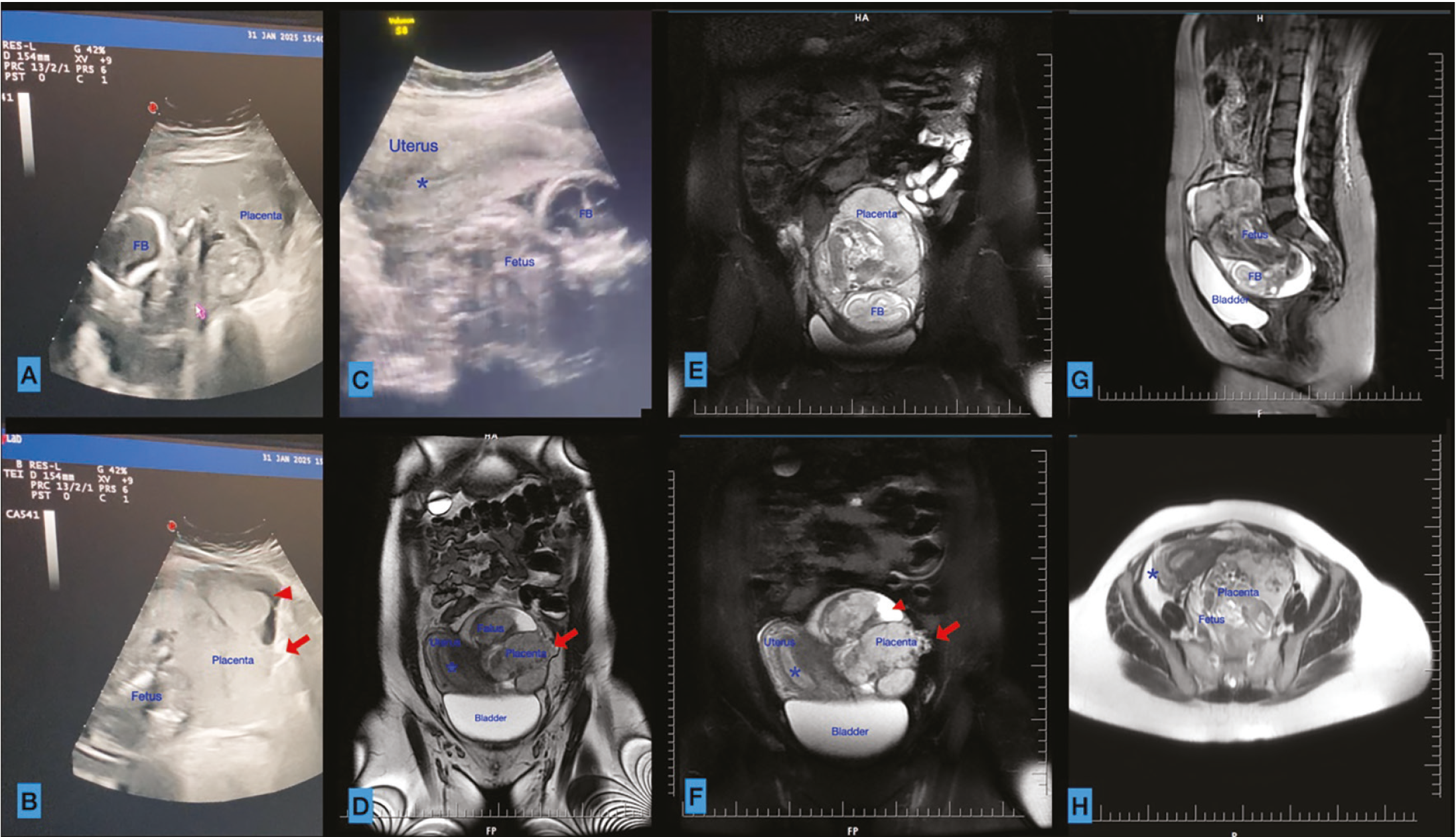
Blue *: Endometrium, red arrowhead: placental lacuna. Red arrow: Invasion of the placenta into surrounding tissues, white arrow: fetus
FB: Fetal brain
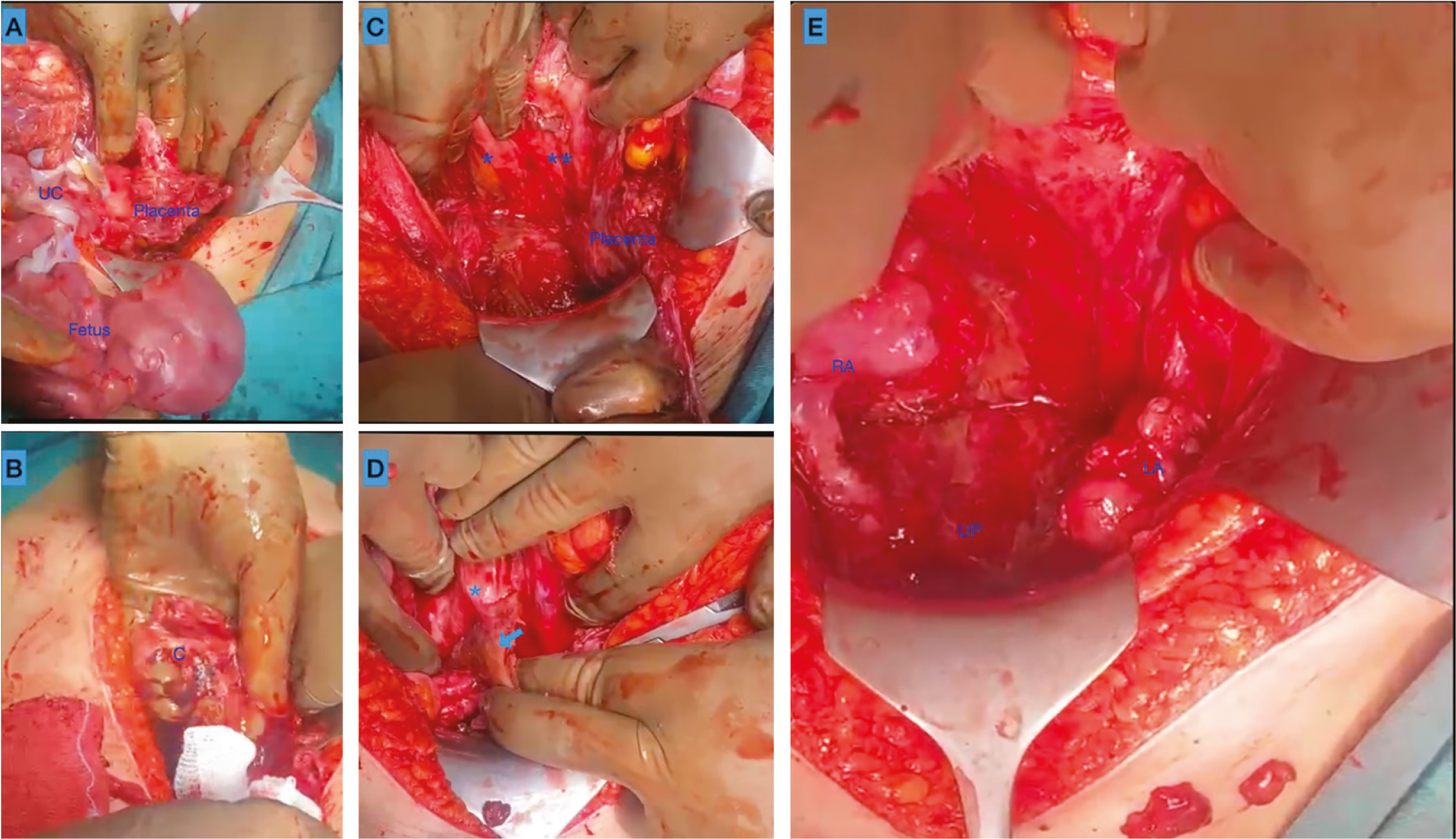
Written and verbal consent was obtained after all risks were explained to the patient. Although fetal cardiac activity was present, immediate laparotomy was preferred because of persistent severe abdominal pain, clinical concern for acute abdomen, and the possibility of progressive maternal risk with advancing gestation. The decision was made after evaluation of the imaging findings and counseling about the risks of hemorrhage, bowel injury, vascular injury, possible organ loss, and fetal loss. The abdomen was opened through a sub-umbilical median incision (). The bladder, omentum, and gestational structures appeared conglomerated and adherent to each other, producing a ‘‘frozen pelvis’’ appearance. The gestational sac was separated from the sigmoid colon, bladder, and omentum by blunt and sharp dissection (). Upon inspection, the placenta was adherent to the peritoneum of the pouch of Douglas, the sigmoid colon, and the parietal peritoneum over the left iliac vessels. Invasion of the iliac vessels was not observed. A female infant weighing 1100 gr, with an Apgar of 0, was delivered from the intra-abdominal cavity. The placenta was removed by dissection from the peritoneum of Douglas, the serosa of the sigmoid colon, the posterior uterus, and the adnexa. The uterus, bilateral ovaries, and fallopian tubes were also preserved. After bleeding was controlled, the abdomen was closed in accordance with the anatomical layers. No blood transfusion was needed. The patient, who did not develop any complications during postoperative follow-up, was discharged with a full recovery. After the patient’s identity was anonymized, written consent was obtained from the patient for the publication of her data and images.
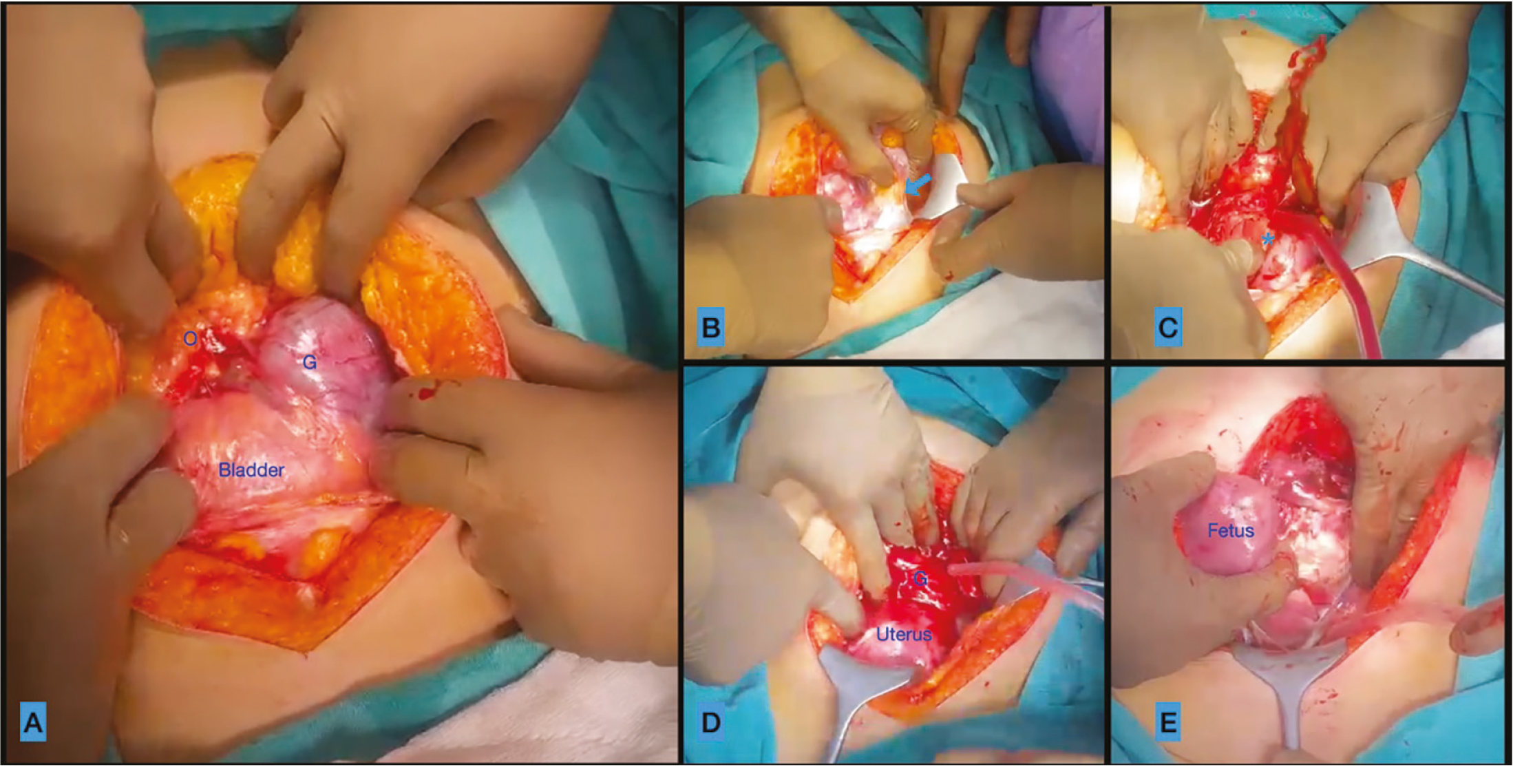
O: Omentum, G: Gestational sac. Blue arrow: Adhesion between sigmoid colon and gestational sac, blue, *: Fetal head
Discussion
Although AAP is rare, its incidence increases in women with risk factors for ectopic pregnancy(). It is known that conditions such as fallopian tube-related diseases (such as ectopic pregnancy, tubal surgery, sterilization and pelvic inflammatory disease), contraception, endometriosis, smoking, history of infertility and intra-abdominal surgery increase the risk(-). In addition, low socioeconomic status, inadequate or incomplete antenatal care, and limited medical facilities may cause delays in the diagnosis of AP and increase the frequency of AAP. In our case, no known risk factors for AAP were identified other than infertility and smoking. However, published reports also show that AAP may occur even in women with access to antenatal care, particularly when symptoms are non-specific or imaging findings are misinterpreted(-).
There are two types of AAP based on their formation. In secondary AP, the pregnancy initially implants in the fallopian tubes. Subsequently, following tubal abortion or rupture, it attaches to the parietal peritoneum and continues to grow. In the less frequent primary AP, the gestational sac is located above any peritoneal surface in the abdominal cavity(). In our case, primary AAP was considered because there were no signs of early-stage pregnancy-related injuries (tuba-utero-peritoneal fistula) in the tubes, ovaries, and uterus. Nevertheless, secondary AP cannot be fully excluded in many advanced cases, because the original implantation site may no longer be clearly identifiable at laparotomy(, ).
AAP may present with clinical presentations ranging from an asymptomatic state to hemorrhagic/septic shock(-). Depending on the location of the gestational sac in the abdomen, patients may present with non-specific symptoms such as nausea, vomiting, abdominal pain, constipation, and ileus. This creates difficulties in making a correct diagnosis. In our case, the symptoms, abdominal pain, constipation, and dyspareunia, which increased in severity over time following delayed menstruation, could be explained by the placement of the fetus in the pouch of Douglas and of the placenta in the rectosigmoid colon. The progressively worsening abdominal pain and tenderness in our patient were clinically significant, as they raised concern for an acute abdomen and influenced the decision for immediate surgical intervention.
It may not always be possible to diagnose AAP(). The diagnosis may be missed in 50-70% of patients due to the presence of non-specific symptoms, suboptimal anatomical evaluation due to advanced gestational age, and technical/structural limitations in imaging(, ). Therefore, the diagnosis of AAP is difficult. The most important parameter in the diagnostic process is the inclusion of AAP in the list of possible diagnoses. Especially in patients who have not undergone any imaging evaluation in the first trimester, as in our case, AAP should be considered in the differential diagnosis. In our case, we suspected an AAP on transabdominal sonography. We established a definitive diagnosis of AAP after transvaginal USG demonstrated empty endocervical and endometrial canals. We then evaluated the relationship between the placental bed and the intestinal and vascular structures using non-contrast-enhanced MRI. MRI was particularly useful in our case because it helped define placental topography and suggested the absence of major iliac vascular invasion, thereby contributing to surgical planning. Similar reports have emphasized that MRI may provide additional value over ultrasonography in delineating placental implantation and adjacent organ relationships in AAP(, , ).
Perinatal mortality in AAP is approximately 72%(). Perinatal mortality may be related to gestational age and inadequate placental perfusion. In our case, the fetus’s gestational age of 24 weeks was considered the main cause of fetal death. The risk of maternal mortality and morbidity also increases with AAP. Although maternal mortality is approximately 12%, abdominal bleeding requiring blood transfusion occurs in 80% of patients(). Additionally, the risks of sepsis, relaparotomy, hysterectomy, oophorectomy, and vascular and intestinal injuries increase. The most important factors determining these risks are the gestational week at the time of diagnosis and the location of the placenta. In our case, although the placenta was located in the peritoneum over the sigmoid colon and iliac vessels, we achieved surgical recovery without complications, organ injury, or loss. One possible reason for this finding is that gestational age was relatively low. As pregnancy progresses, invasion may deepen, and treatment may become more difficult. This possibility was one of the reasons we did not favor prolonging pregnancy in the presence of severe pain and acute abdominal findings.
Management of AAP is difficult and requires care at an experienced tertiary care hospital. Evidence-based treatment is not available because it is rare. Treatment should be determined by considering the condition of the fetus at the time of diagnosis, the week of pregnancy, and the relationship of the placenta to the intra-abdominal organs. When the fetus is non-viable or has an anomaly at diagnosis, necessary preparations can be made and surgery performed immediately. However, it may not always be possible to make this decision when the fetus is alive or healthy. There are cases in the literature where, after diagnosis, prolonging the pregnancy is offered as an option with patient consent to ensure fetal maturation(). At the same time, expectant management has generally been described in carefully selected stable patients under close inpatient surveillance and multidisciplinary monitoring(-). In our patient, despite fetal cardiac activity, immediate laparotomy was preferred because of persistent severe abdominal pain, concern for an acute abdomen, and the possibility of increasing maternal risk with advancing gestation.
Since the MRI examination revealed that the placenta was located away from the main vessels and intestines, no problems may have occurred during the removal of placental tissues in this case. However, in cases in which placental invasion involves the major vascular structures or the bowel, any delay may increase the complication rate. In the present case, the sigmoid colon and iliac vessels may be at risk of deeper involvement during advanced gestational weeks. Therefore, the decision to proceed with immediate surgery in our case was based not only on fetal status but primarily on maternal symptoms and the anticipated progression of local invasion.
In the literature, it has been emphasized that treatment alternatives such as leaving the placenta in place and postoperative arterial embolization or methotrexate administration can be successfully applied in surgical treatment because removing the placenta during surgery may cause excessive bleeding and organ damage/loss in the mother(, -). When the placenta cannot be removed, close, long-term follow-up is required because resorption of the placenta at the site of invasion may lead to sepsis and secondary bleeding. Recent reports have also shown that even embolization-assisted conservative management may be followed by abscess formation, infection, or delayed reoperation(, ). Conversely, attempts at placental removal may result in catastrophic hemorrhage, shock, relaparotomy, or hysterectomy in anatomically unfavorable cases(, ). There are also cases in the literature in which the placenta was removed and treated successfully(, , -). In the present case, the placenta was removed without any complications. Taken together, these findings suggest that placental management should be individualized according to placental location, expected vascularity, adjacent organ involvement, maternal condition, and local surgical expertise, rather than following a single universal strategy(, , , ).
As summarized in the comparative literature table (), published reports do not support a single standard approach to AAP. Instead, management appears to depend on the interaction between maternal symptoms, fetal viability, placental anatomy, and institutional preparedness(, , , , , ). Our case adds to the limited literature suggesting that, in selected symptomatic patients without major vascular invasion on imaging, MRI-guided immediate organ-sparing surgery with placental removal may be feasible(, ).
Table 1. Comparative summary of published advanced abdominal pregnancy cases and case series relevant to diagnostic and management dilemmas
Study | GA at diagnosis | Maternal/fetal status at diagnosis | Imaging | Management | Placental management | Maternal outcome | Fetal/neonatal outcome |
|---|---|---|---|---|---|---|---|
Present case/Türkiye | 24 w | Stable mother; live fetus | US + MRI | Immediate surgery | Removed | Uneventful recovery | 1100 g female, Apgar 0 |
Kim et al.(), 2013/Korea | 18 w | Stable mother; live fetus | US + MRI | Expectant to 34 w | Removed | Uneventful; uterus preserved | Live female, 2100 g |
Marcellin et al.(), 2014/France | 22 w | Stable asymptomatic inpatient; live fetus | US + MRI | Expectant to 32 w | Left in situ + embolization/follow-up | Maternal survival | Live birth |
Harirah et al.(), 2016/USA | 23 w and 28 w | Stable mothers; live fetuses | US + MRI | Individualized: planned delivery vs. expectant management | Partial removal in one; in situ in one | Both survived | Favorable neonatal outcomes |
Muroni et al.(), 2021/Burundi | 37 w | Stable mother; live fetus | US | Elective laparotomy | Removed | Massive hemorrhage, shock, relaparotomy; survived | Live birth; later infant death |
Owie et al.(), 2022/Nigeria | 27 w | Septic mother; live fetus | US | Immediate surgery | Partially left in situ | Survived; transfusion required | Neonatal death within 24 h |
Legesse et al.(), 2023/Ethiopia | 37 + 6 w | Stable mother; live fetus | Expert US | Immediate surgery | Removed | No maternal complication | Live female, 2060 g; foot deformity |
Alwafai et al.(), 2024/Germany | Late, missed until IUFD | Deteriorating mother; IUFD | US | Failed induction, then urgent laparotomy | Removed | Good postoperative recovery | Non-viable fetus |
Emmanuel et al.(), 2026/Ghana | 29 + 3 w | Stable mother; live fetus | US | Expectant to 32 w | Removed | Hemorrhage, transfusion, postoperative abscess/relaparotomy; survived | Live male, 1.6 kg, no gross anomaly |
Sunday-Adeoye et al.(), 2011/Nigeria | Mean 32.5 w (range 27-38 w) | Variable maternal status; mixed fetal outcomes | Limited imaging | Mixed surgical management | Removed in 11; in situ in others | 1 maternal death | Poor perinatal outcomes overall |
Harries et al.(), 2025/South Africa | Median 31 + 4 w | Variable maternal status; 12 live births | US ± MRI | Mostly expedited delivery within 48 hours | Variable | 2/17 maternal deaths | 12 live births; 10 discharged home |
Hoshino et al.(), 2021/Japan | 20-42 w (median 37.5 w) | Variable maternal status; mostly live births in later cases | US + MRI in recent cases | Mixed | Removed at laparotomy in 5/10; delayed removal in others | Maternal survival emphasized | 6/7 live births had mainly compression-related deformities |
Çetin Arslan(), 2025/Türkiye | 20 w and 29 w | Both diagnosed with live fetuses | US + MRI | Immediate surgery in both | Removed in both | Both survived; high blood loss/ICU care | Both live births |
GA: Gestational age, US: Ultrasonography, MRI: Magnetic resonance imaging, ICU: Intensive care unit, w: Weeks, IUFD: Intrauterine fetal death
Conclusion
AAP is a preventable yet life-threatening condition associated with high maternal and fetal morbidity and mortality. Early diagnosis—particularly during the first trimester—is critical to improving outcomes. Surgical risk is largely dependent on the location and extent of placental invasion. While placental removal carries significant hemorrhagic risk, leaving the placenta in situ may result in secondary complications. MRI provides valuable information on placental localization and potential vascular involvement and may be particularly helpful for preoperative planning for suspected AAP cases. Management should occur in tertiary centers with multidisciplinary teams, and each case should be approached individually, with informed patient consent and detailed surgical planning. In symptomatic patients with severe pain or acute abdominal findings, immediate surgery may be justified despite the presence of fetal cardiac activity. In selected cases without major vascular invasion, organ-sparing placental removal may be feasible.
Footnotes
Ethics
Informed Consent: After the patient’s identity was anonymized, written consent was obtained from the patient for the publication of her data and images.
Authorship Contributions
Surgical and Medical Practices: M.B., P.D., G.K.A., Concept: M.B., G.K.A., Design: M.B., P.D., Data Collection or Processing: M.B., P.D., G.K.A., Analysis or Interpretation: M.B., P.D., G.K.A., Literature Search: M.B., Writing: M.B., P.D., G.K.A.
Conflict of Interest: No conflict of interest was declared by the authors.
Financial Disclosure: The authors declared that this study received no financial support.
References
Panelli DM, Phillips CH, Brady PC. Incidence, diagnosis and management of tubal and nontubal ectopic pregnancies: a review. Fertil Res Pract. 2015;1:15.
Chauhan SP, Bhalwal AB. Abdominal pregnancy. Simpson LL, Chakrabarti A, eds. UpToDate; 2025. Available from: https://www.uptodate.com/contents/abdominal-pregnancy?search=Abdominal%20pregnancy&source=search_result&selectedTitle=1%7E132&usage_type=default&display_rank=1
Muroni M, Butoyi JMV, Shimirimana M, Mulemangabo M, Nkurunziza J, Caravaggi P. Hemoperitoneum during removal of the placenta in advanced abdominal pregnancy with live fetus delivered at 37 weeks of gestation. A case report in a low-resource setting and literature review. Int J Surg Case Rep. 2021;80:105694.
Rohilla M, Joshi B, Jain V, Neetimala, Gainder S. Advanced abdominal pregnancy: a search for consensus. Review of literature along with case report. Arch Gynecol Obstet. 2018;298:1-8.
Harries S, Tooke L, Imbeault JC, Matjila M, Pillay S. Advanced abdominal pregnancy at a tertiary hospital in South Africa: a case series. AJOG Glob Rep. 2025;5:100500.
Sunday-Adeoye I, Twomey D, Egwuatu EV, Okonta PI. A 30-year review of advanced abdominal pregnancy at the Mater Misericordiae Hospital, Afikpo, southeastern Nigeria (1976-2006). Arch Gynecol Obstet. 2011;283:19-24.
Hoshino T, Mori T, Fujii Y, Yoshioka S. Advanced abdominal pregnancy (AAP) after 20 weeks of gestation in Japan: a retrospective review. Obstet Gynecol Int. 2021;2021:6624404.
Li C, Zhao WH, Zhu Q, Cao SJ, Ping H, Xi X, et al. Risk factors for ectopic pregnancy: a multi-center case-control study. BMC Pregnancy Childbirth. 2015;15:187.
Zhang D, Shi W, Li C, Yuan JJ, Xia W, Xue RH, et al. Risk factors for recurrent ectopic pregnancy: a case-control study. BJOG. 2016;123(Suppl 3):82-9.
Watrowski R, Lange A, Möckel J. Primary omental pregnancy with secondary implantation into posterior cul-de-sac: laparoscopic treatment using hemostatic matrix. J Minim Invasive Gynecol. 2015;22:501-3.
Alwafai Z, Kolbe C, Kruse-Wieczorek J, Khanji MN, Zygmunt M. Challenging diagnosis of late abdominal pregnancy: a case study of misdiagnosis and fetal death in the third trimester. Am J Case Rep. 2024;25:e943625.
Nassali MN, Benti TM, Bandani-Ntsabele M, Musinguzi E. A case report of an asymptomatic late term abdominal pregnancy with a live birth at 41 weeks of gestation. BMC Res Notes. 2016;9:31.
Chen Y, Peng P, Li C, Teng L, Liu X, Liu J, et al. Abdominal pregnancy: a case report and review of 17 cases. Arch Gynecol Obstet. 2023;307:263-74.
Ranaei-Zamani N, Palamarchuk T, Kapoor S, Kaler MK, Atueyi F, Allen R. Diagnostic challenges of an abdominal pregnancy in the second trimester. Case Rep Obstet Gynecol. 2021;2021:7887213.
Kim MJ, Bae JY, Seong WJ, Lee YS. Sonographic diagnosis of a viable abdominal pregnancy with planned delivery after fetal lung maturation. J Clin Ultrasound. 2013;41:563-5.
Marcellin L, Ménard S, Lamau MC, Mignon A, Aubelle MS, Grangé G, et al. Conservative management of an advanced abdominal pregnancy at 22 weeks. AJP Rep. 2014;4:55-60.
Ramphal S, Khaliq OP, Abel T, Moodley J. Expectant management of advanced abdominal pregnancies: is it justifiable? Eur J Obstet Gynecol Reprod Biol. 2023;281:99-108.
Valenzano M, Nicoletti L, Odicino F, Cocuccio S, Lorenzi P, Ragni N. Five-year follow-up of placental involution after abdominal pregnancy. J Clin Ultrasound. 2003;31:39-43.
Oneko O, Petru E, Masenga G, Ulrich D, Obure J, Zeck W. Management of the placenta in advanced abdominal pregnancies at an East african tertiary referral center. J Womens Health (Larchmt). 2010;19:1369-75.
Joshi RR. Experience of Secondary abdominal pregnancy reported at a rural tertiary care center of Western India: a case series. Cureus. 2024;16:e55663.
Lee C. Abdominal pregnancy in a low-resource setting. Obstet Gynecol. 2015;125:1039-1.
El-Agwany AS, El-Badawy el-S, El-Habashy A, El-Gammal H, Abdelnaby M. Secondary advanced abdominal pregnancy after suspected ruptured cornual pregnancy with good maternal outcome: a case with unusual gangrenous fetal toes and ultrasound diagnoses managed by hysterectomy. Clin Med Insights Womens Health. 2016;9:1-5.
Legesse TK, Ayana BA, Issa SA. Surviving fetus from a full term abdominal pregnancy. Int Med Case Rep J. 2023;16:173-8.
Emmanuel OA, Adu RB, Walid RM, Tutu ACO, Albert AA, Fadila AM, et al. Expectant management of advanced abdominal pregnancy with successful outcome: a case report and literature review. Womens Health (Lond). 2026;22:17455057261446946.
Çetin Arslan H. Advanced abdominal pregnancy case series successfully treated with laparotomy. Turkiye Klinikleri J Case Rep. 2025;33:65-8.
Harirah HM, Smith JM, Dixon CL, Hankins GD. Conservative management and planned surgery for periviable advanced extrauterine abdominal pregnancy with favorable outcome: report of two cases. AJP Rep. 2016;6:e301-8.
Owie E, Lewu A, Anumni C. A case report of an advanced abdominal pregnancy with sepsis. J Obstet Gynaecol. 2022;42:1556- 7.