
Abstract
This paper presents a comprehensive cadaveric dissection atlas detailing the retroperitoneal anatomy essential to advanced pelvic surgery. Developed by a multidisciplinary team of anatomists, gynecologists, and gynecologic oncologists, the study provides a systematic, layer-by-layer guide for navigating the pelvic avascular spaces. It details five critical surgical corridors: the presacral (retrorectal) space, the pararectal space (divided into the Latzko and Okabayashi compartments), the paravesical space (medial and lateral compartments), the prevesical (retropubic/Retzius) space, and the medial psoas space (laterovascular plane). This atlas emphasizes the use of reliable landmarks, such as the sacral promontory, the obliterated umbilical artery, and the ureter, to define anatomical boundaries and ensure safe surgical practice. The primary objective is to maintain adequate exposure after retroperitoneal entry and to perform layer-by-layer surgical dissection to identify critical anatomical structures. The superior hypogastric plexus; hypogastric nerve; pelvic splanchnic nerves; inferior hypogastric plexus with its vesical and rectal branches; the internal iliac artery with its posterior and anterior trunk branches (including the superior gluteal, iliolumbar, lateral sacral, uterine, inferior gluteal, pudendal, obturator, middle rectal, and inferior and superior vesical arteries, and the obliterated umbilical artery); the external iliac artery; and the corresponding internal and external iliac veins were discussed. Additionally, the somatic nerves, obturator nerve, lumbosacral trunk, sacral nerves, sciatic nerve, genitofemoral nerve, and femoral nerve were reviewed. The parietal fascial planes, pubocervical fascial structure, and visceral compartments were evaluated as part of the whole. Respecting fascial planes—particularly the presacral fascia—is mandatory to avoid catastrophic hemorrhage and autonomic nerve injury. These spaces serve as the “neurovascular roadmap” for complex procedures, including radical hysterectomy, nerve-sparing pelvic surgery, pelvic lymphadenectomy, and hemorrhage control. Mastery of these interconnected retroperitoneal compartments facilitates a transition from organ-based to space-oriented surgery, significantly reducing morbidity while maintaining oncologic radicality.
Keywords: Retroperitoneum, pelvic avascular spaces, ureter, internal iliac artery, nerve-sparing surgery
Introduction
The pelvic bones (sacrum posteriorly, pubic bone anteriorly, ilium posterolaterally on the superior side, ischium anterolaterally on the inferior side) encircle the pelvic structures. The inner layer of the pelvic bones is formed by the muscles (obturator internus laterally and levator ani at the base) and ligaments, all of which are covered by the parietal fascia. The loose, connective, and supportive fatty tissue surrounds the midline pelvic organs (the rectum posteriorly, the bladder anteriorly, and the uterus between them), and this compartment is covered by the parietal peritoneum(). This review focuses on the detailed surgical anatomy of the pelvic retroperitoneum as encountered during incision of the pelvic parietal peritoneum and layer-by-layer dissection from superficial to deep structures. The pelvic avascular spaces—presacral, pararectal, paravesical, and prevesical—and the laterovascular plane (medial psoas space) are well described in the literature.
Methods
This educational cadaveric dissection was performed at the Anatomy Department Laboratory of the University of Health Sciences Türkiye, Gülhane Faculty of Medicine, Ankara, Türkiye. The multidisciplinary team, consisting of anatomists, gynecologists, and gynecologic oncologists, worked collaboratively. An educational video lecture of this video can be found on the YouTube platform (https://www.youtube.com/watch?v=x_VmYaQxclM&t=1402s).
Presacral Space (Retrorectal Space) Definition and Surgical Concept
The presacral space is a midline, retroperitoneal, avascular pelvic space located anterior to the sacrum and posterior to the rectum, extending from the pelvic brim (sacral promontory) to the pelvic floor. It represents the central posterior pelvic dissection plane and serves as a critical corridor for posterior pelvic surgery, nerve identification, and en bloc resections in gynecologic oncology. The presacral space is particularly important for the orientation of the superior hypogastric plexus, hypogastric nerves, and major pelvic vessels, and it acts as the anatomical bridge between the paraaortic region and the posterior pelvic compartments.
Anatomical Boundaries ( and )
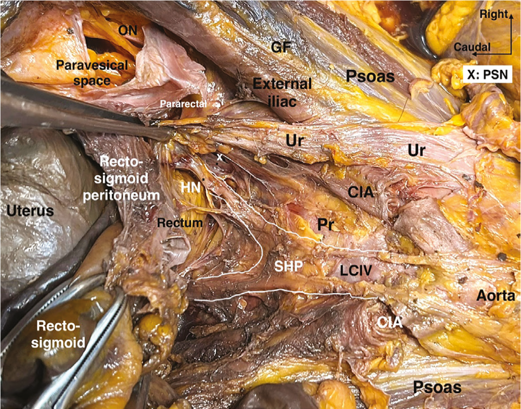
CIA: Common iliac artery, Ur: Ureter, LCIV: Left common iliac vein, Pr: Promontorium, SHP: Superior hypogastric plexus, HN: Hypogastric nerve, ON: Obturator nerve, PSN: Pelvic splanchnic nerves, GF: Genitofemoral nerve
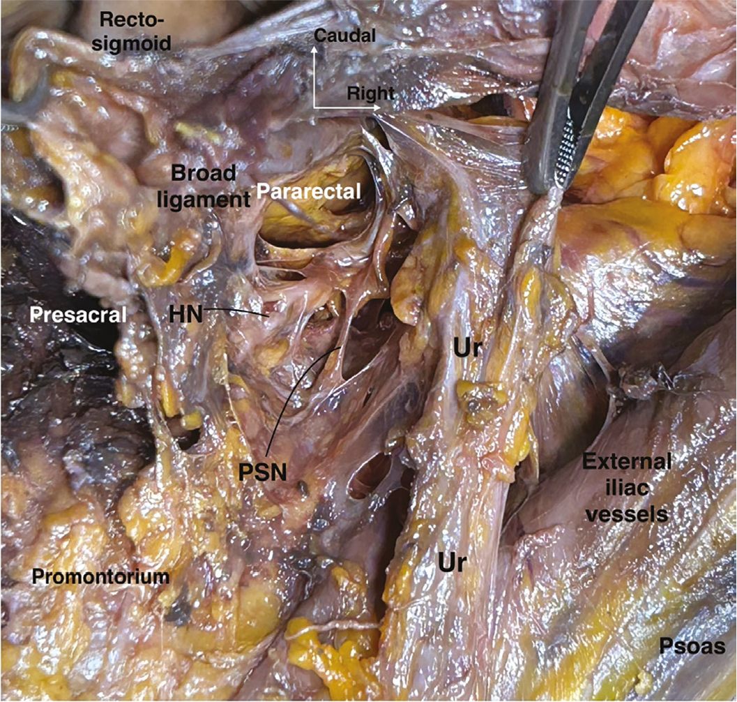
Ur: Ureter, HN: Hypogastric nerve, PSN: Pelvic splanchnic nerves
The presacral space is defined by constant and easily identifiable landmarks:
• Anterior boundary:
Rectum,
Mesorectal fascia (mesorectum).
• Posterior boundary:
Sacrum,
Presacral fascia (Waldeyer’s fascia).
• Lateral boundaries:
Ureters,
Common iliac artery, internal iliac artery (proximal portions).
• Superior boundary:
Pelvic brim (sacral promontory),
Inferior border of the sigmoid mesocolon.
• Inferior boundary:
Levator ani muscle,
Anococcygeal ligament.
These boundaries position the presacral space as the deepest posterior pelvic avascular plane, in close proximity to major vascular and autonomic neural structures.
Surgical Development (Step-by-Step)
1. Identification of the Sacral Promontory (Promontorium)
The sacral promontory is the key landmark for safe entry.
It marks the transition between the paraaortic and pelvic retroperitoneum.
2. Peritoneal Incision
The parietal peritoneum is incised:
• On the right, inferior to the radix mesenterii of the small bowel,
• On the left, along the Toldt’s line after mobilization of the sigmoid colon.
3. Recognition of Critical Structures
Ureters are identified laterally.
The left common iliac vein is visualized anterior to the lumbar 5th vertebra, superior to the sacral promontory.
Superior hypogastric plexus is identified anterior to the left common iliac vein or lumbar 5th vertebra, attached to the retrorectal adipose tissue (posterior mesorectal plane).
4. Blunt Dissection
Dissection proceeds anterior to the presacral fascia.
Sharp dissection posterior to the presacral fascia must be avoided to prevent massive venous bleeding.
5. Caudal Extension
The space is deepened toward the pelvic floor.
The superior hypogastric plexus is divided into right and left hypogastric nerves.
Hypogastric nerves are followed caudally and caudolaterally at the anterior (anterolateral) part of the sacrum towards the pararectal spaces.
Proper development results in a bloodless midline surgical corridor that allows safe posterior pelvic mobilization.
Contents and Key Structures
Although the presacral space is considered avascular, its walls are closely associated with critical elements:
• Neural
Superior hypogastric plexus at the superior part, mixed nerve with dominantly sympathetic and also partially parasympathetic activity.
Bilateral hypogastric nerve lies caudolaterally at the anterolateral part of the sacrum and the posterolateral part of the rectum.
• Vascular
Median sacral artery and vein, posterior to presacral fascia.
Left common iliac vein, superiorly.
Common iliac artery, laterally.
• Fascial
Presacral (Waldeyer’s) fascia, the posterior boundary, covering the sacrum.
• Lymphatic
Presacral lymphatic tissue (limited but clinically relevant).
Understanding these contents is essential to avoid catastrophic hemorrhage and autonomic nerve injury.
Surgical Applications
The presacral space has multiple critical applications in pelvic surgery:
• Radical hysterectomy
Provides a posterior access and dissection plane.
Enables safe identification and preservation of the superior hypogastric plexus and hypogastric nerve.
• Nerve-sparing surgery
Serves as the primary orientation plane for autonomic nerve preservation, the superior hypogastric plexus.
• Posterior pelvic resections
Essential for rectosigmoid mobilization.
Facilitates posterior or total pelvic exenteration.
• Ovarian cancer cytoreduction
Enables en bloc resection of the uterus, rectum, and sigmoid colon in cases of obliterated Douglas pouch.
• Hemorrhage control
Provides access to proximal vascular control when pelvic bleeding is encountered.
These applications emphasize the presacral space as a strategic posterior pelvic corridor rather than a simple anatomical cavity.
Key Surgical Insight
The presacral space is the anatomical gateway to the pelvic autonomic nervous system and to the identification of the ureter at the pelvic brim level. Respecting the presacral fascia is the single most important rule to prevent uncontrollable bleeding and nerve injury.
Pararectal Space
Definition and Surgical Concept
The pararectal space is a paired, retroperitoneal, avascular pelvic space located lateral to the rectum and medial to the internal iliac vascular system. It represents a critical surgical corridor within the posterior pelvic compartment, providing access to the uterosacral ligament, pelvic autonomic nerves, internal iliac vessels, and posterior (dorsal) parametrium. In gynecologic oncology, the pararectal space is essential for radical hysterectomy, nerve-sparing procedures, and posterior pelvic resections, serving as a key orientation space for differentiating vascular from neural planes.
Anatomical Boundaries ( and )
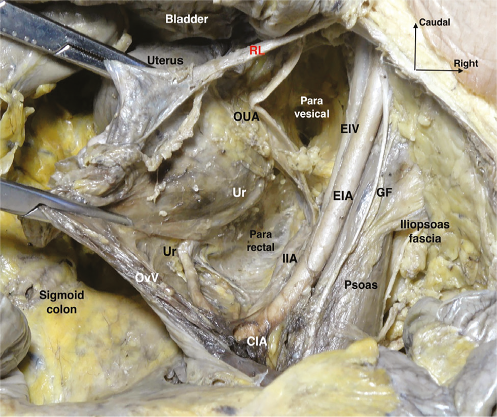
CIA: Common iliac artery, EIA: External iliac artery, EIV: External iliac vein, IIA: Internal iliac artery, GF: Genitofemoral nerve, RL: Round ligament, OvV: Ovarian vessels, OUA: Obliterated umbilical artery, Ur: Ureter
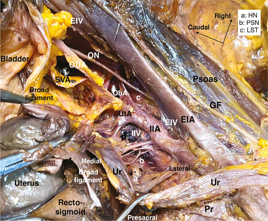
EIA: External iliac artery, EIV: External iliac vein, IIA: Internal iliac artery, IIV: Internal iliac vein, GF: Genitofemoral nerve, HN: Hypogastric nerve, LST: Lumbosacral trunk, ObA: Obturator artery, ON: Obturator nerve, OUA: Obliterated umbilical artery, Pr: Promontory, PSN: Pelvic splanchnic nerves, SVA: Superior vesical artery, Ur: Ureter, UtA: Uterine artery
The pararectal space is anatomically defined by the relationship between the rectum, ureter, and internal iliac system:
• Medial boundary:
Rectum,
Uterosacral ligament (posterior parametrium, rectouterine ligament/ligamentum rectouterinum),
Ureter, which divides the pararectal space into medial and lateral compartments.
• Lateral boundary:
Internal iliac artery and its branches (internal iliac vein is noticed inferior to the internal iliac artery).
• Anterior boundary:
Cardinal ligament (lateral parametrium, parauterine and paracervix tissue),
Uterine artery-vein complex with the pelvic autonomic nerves.
• Posterior boundary:
Sacrum,
Presacral fascia.
• Superior boundary:
Pelvic parietal peritoneum.
• Inferior boundary:
Levator ani muscle (iliococcygeus component).
These boundaries position the pararectal space at the crossroads of the vascular, neural, and visceral components of pelvic anatomy, making it one of the most critical pelvic spaces when approaching the frozen pelvis.
Subdivision of the Pararectal Space ()
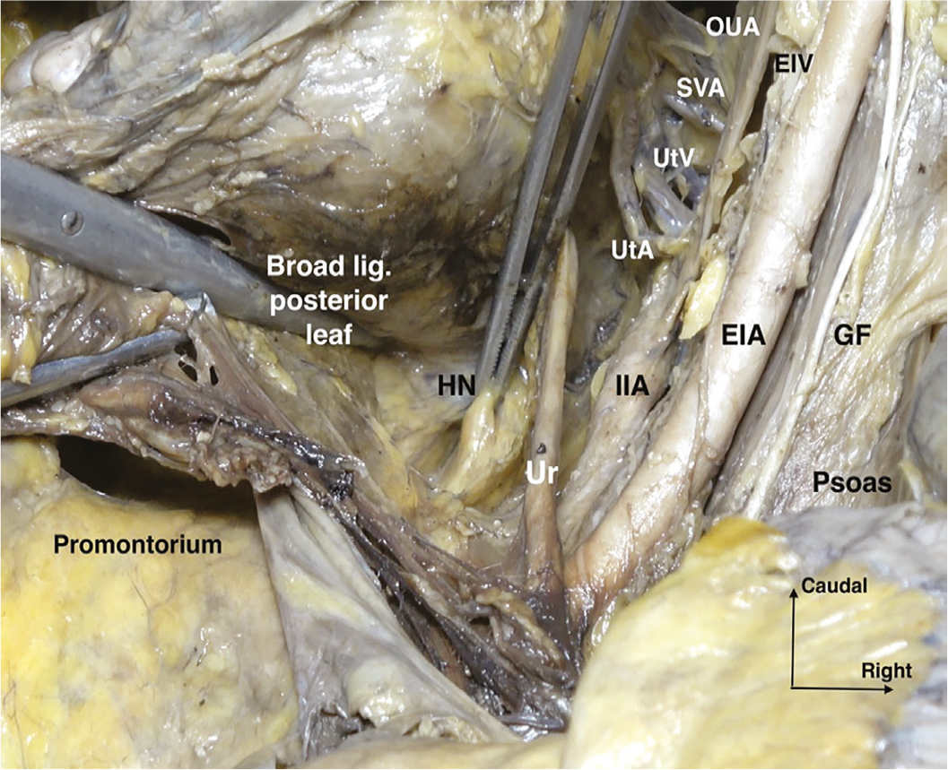
GF: Genitofemoral nerve, EIA: External iliac artery, EIV: External iliac vein, IIA: Internal iliac artery, Ur: Ureter, HN: Hypogastric nerve, UtA: Uterine artery, UtV: Uterine vein, SVA: Superior vesical artery, OUA: Obliterated umbilical artery, lig: Ligament
If the ureter is dissected and lateralized from the posterior leaf of the broad ligament, it is the key landmark dividing the pararectal space into two surgically distinct compartments:
1. Lateral pararectal space (Latzko space)
Located between the ureter (medially) and the internal iliac artery (laterally),
Primarily vascular in orientation, following the internal iliac artery will lead to the uterine artery, which is the first anteromedial branch arising from the internal iliac artery,
Contains the origin and proximal parts of the pelvic splanchnic nerves.
2. Medial pararectal space (Okabayashi space)
Located between the ureter (laterally) and the rectum/uterosacral ligament (medially),
Contains critical pelvic autonomic nerve structures, the hypogastric nerve, caudal (medial distal) parts of the pelvic splanchnic nerves, and the inferior hypogastric plexus,
This subdivision is fundamental to nerve-sparing surgical procedures (radical hysterectomy, endometriosis surgery, mesorectal resection) because neural elements are concentrated in the medial compartment.
Surgical Development (Step-by-Step)
1. Retroperitoneal entry
Achieved by incising the lateral parietal peritoneum:
• Cut the pelvic lateral parietal peritoneum, along and lateral to the infundibulopelvic ligament (ovarian vessels),
• Or medial approach, medial to the infundibulopelvic ligament, cut the posterior leaf of the broad ligament, and notice that the ureter is attached to the posterior leaf of the broad ligament inferior to the ovarian vessels.
2. Identification of the ureter
The ureter is identified at the pelvic brim, crossing over the common iliac vessels,
It is followed caudally along the posterior leaf of the broad ligament, inferior to the ovarian vessels.
3. Creation of the lateral pararectal space (Latzko)
Blunt dissection between the ureter and the internal iliac artery,
Develops a relatively avascular plane anterior to the sacrum.
4. Development of the medial pararectal space (Okabayashi)
After mobilization or lateralization of the ureter from the broad ligament posterior leaf,
Dissection proceeds between the ureter and rectum/uterosacral ligament.
5. Caudal maturation
The space deepens toward the pelvic floor and the levator ani.
Notice the internal iliac vein inferior (inferior in surgical supine position, posterior in anatomical position) to the internal iliac artery.
The hypogastric nerve, at the medial pararectal space, inferior to the ureter and attached to the perirectal visceral fascia, becomes identifiable (sympathetic activity). The ureter and the hypogastric nerve lie within the same fascial sheet, the ureterohypogastric fascia.
The pelvic splanchnic nerves, located in the lateral pararectal space at the inferomedial aspect of the internal iliac vein, arise from the sacral 2-4 foramina (parasympathetic activity). They lie obliquely medial to the paracervix and are attached to the paracervix inferior to the deep uterine or vaginal vein.
The hypogastric nerve and the pelvic splanchnic nerves merge to form the inferior hypogastric plexus at the medial part of the paracervix, inferior to the level of the (deep) uterine vein, which is a mixed ganglion with parasympathetic and sympathetic innervation.
Correct development creates a bloodless, anatomically stratified space, permitting precise oncologic and nerve-preserving dissection.
Contents and Key Structures
Although avascular, the pararectal space is closely related to critical structures:
• Neural
Hypogastric nerve, medially,
Pelvic splanchnic nerves, laterally,
Inferior hypogastric plexus, at the medial caudal compartment, closely related to the medial paracervix, lateral to the rectum and posterolateral to the upper vagina.
• Vascular
Internal iliac artery branches, in the lateral compartment,
Uterine artery, at the anterior part, superior to the ureter,
Deep uterine vein, at the anterior part, inferior to the ureter.
• Fascial
Uterosacral ligament, medially,
Presacral fascia, posteriorly.
• Visceral
Rectum, medially,
Ureter, central landmark.
• Beyond the pelvic brim, the ureter lies caudomedially within the medial aspect of the pararectal space, is situated inferior to the ovarian vessels, and is attached to the posterior leaf of the broad ligament. Between the pararectal and paravesical spaces, the ureter lies medially, interposed between the parauterine tissue—comprising the uterine artery and associated lymphatic structures (parauterine lymphatic tissue, PULT) located superior to the ureter—and the paracervix, which contains the (deep) uterine vein and related lymphatic tissue, along with the distal segments of the pelvic splanchnic nerves. Following this junction, the ureter traverses the ureteric tunnel in the lateral aspect of the upper vagina, lying between the vesicouterine ligament superiorly and the vesicovaginal ligament inferiorly. Subsequently, the ureter enters the bladder at the trigone. The vesicouterine ligament contains cervicovesical vessels, whereas the vesicovaginal ligament lies adjacent to the vesicovaginal venous plexus and bladder nerve branches. Moreover, the vesicovaginal ligament is continuous with the lateral paracervix.
Recognition of these structures is mandatory to avoid injury to the autonomic nerves and to prevent pelvic organ dysfunction.
Surgical Applications
The pararectal space is indispensable in advanced pelvic surgery.
• Radical hysterectomy
Enables posterior and lateral parametrial resection,
Essential for Querleu-Morrow type B and C procedures.
• Nerve-sparing surgery and ureteric dissection
Allows identification and preservation of the hypogastric nerve and inferior hypogastric plexus with the ureteric dissection and mobilization,
The pelvic splanchnic nerves can be identified and dissected from the paracervix.
• Pelvic lymphadenectomy
Provides orientation for obturator and internal iliac nodal dissection.
• Posterior pelvic resections
Facilitates rectosigmoid mobilization,
Essential in posterior or total pelvic exenteration.
• Complex pelvic and oncologic surgery
Crucial when the rectouterine (Douglas) pouch is obliterated by tumor or fibrosis.
• Hemorrhage control
Provides access to proximal vascular control (internal iliac and uterine arterty) when pelvic bleeding is encountered,
These applications underline the role of the pararectal space as a surgical neurovascular roadmap.
Paravesical Space
Definition and Surgical Concept
The paravesical spaces are bilateral, avascular, retroperitoneal spaces situated lateral to the urinary bladder within the pelvis. It constitutes an essential surgical corridor in the anterior pelvic compartment, facilitating secure access to the lateral pelvic wall, bladder, and parametrial structures. In gynecologic oncology surgery, the paravesical space serves as a key landmark for pelvic lymphadenectomy, radical hysterectomy, nerve-sparing procedures, and hemorrhage management.
Anatomical Boundaries ( and )
The paravesical space is best understood in relation to the bladder medially and the pelvic sidewall laterally.
• Medial boundary:
Urinary bladder,
Pubocervical ligament,
Obliterated umbilical artery (OUA), which divides the paravesical space into medial and lateral compartments.
• Lateral boundary:
The external iliac vessels are observed along the medial aspect of the psoas major muscle. The external iliac artery lies medial to the psoas major muscle, and the external iliac vein lies inferomedial to the artery. The caudal (distal) ends of these vessels constitute the lateral boundary of the paravesical space.
Obturator internus muscle and fascia.
• Anterior boundary:
Superior pubic ramus, pubic bone,
Retropubic (prevesical) space lateral continuity.
• Posterior boundary:
Cardinal ligament [lateral parametrium, parauterine tissue (uterine artery) and paracervix tissue (uterine vein and pelvic autonomic nerves)],
Proximal internal iliac artery branches.
• Inferior boundary:
Pelvic floor (levator ani muscle, pubococcygeus and iliococcygeus).
• Superior boundary:
Pelvic parietal peritoneum, broad ligament anterior leaf,
These boundaries place the paravesical space at the intersection of vascular, neural, and lymphatic surgical planes, emphasizing its importance in oncologic dissection.
Subdivision of the Paravesical Space ()
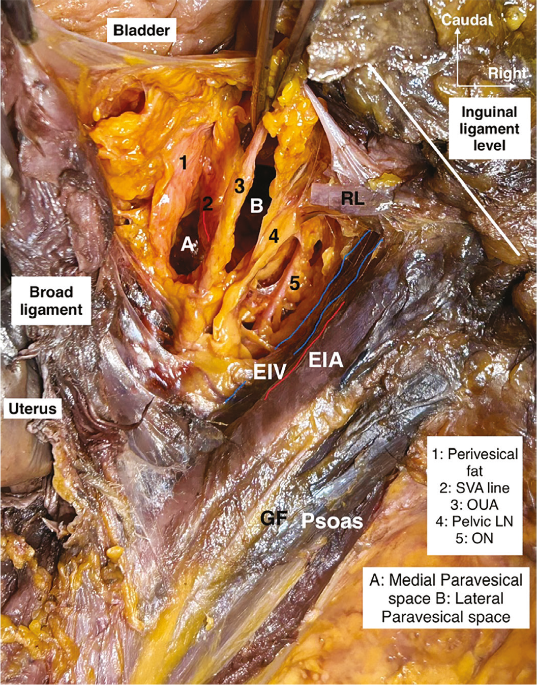
GF: Genitofemoral nerve, EIA: External iliac artery, EIV: External iliac vein, RL: Round ligament, ON: Obturator nerve, LN: Lymph node, OUA: Obliterated umbilical artery, SVA: Superior vesical artery
If the OUA is dissected and lateralized from the perivesical visceral fascia and fatty tissue (the umbilicovesical fascia), it is the key landmark dividing the paravesical space into two distinct compartments:
1. Medial paravesical space
Located between the bladder and the OUA,
Closely related to the anterior (ventral) parametrium and bladder vascular supply.
2. Lateral paravesical space
Located between the OUA and the external iliac vessels,
Continuous laterally with the obturator fossa,
This subdivision is surgically critical because pelvic lymph node dissection is performed through the lateral paravesical compartment, whereas nerve-sparing parametrial dissection of the anterior parametrium is performed through the medial paravesical compartment.
Obturator Fossa
The obturator fossa represents the lateral extension, at the inferior aspect of the external iliac vein, of the lateral paravesical space. Its lateral boundary is delineated by the obturator internus muscle, with the obturator nerve positioned medially relative to the obturator lymph nodes. The obturator nerve exits the pelvis via the obturator canal, located at the superolateral portion of the obturator foramen. The superior margin of the obturator foramen corresponds to the superior pubic ramus. The pelvic anastomotic branches between the obturator vessels and either the inferior epigastric or external iliac vessels are called Corona Mortis vessels (pubic vessels) and are situated over the lateral aspect of the superior pubic ramus. These vessels are predominantly venous anastomoses, with their branches extending from the medial side of the obturator nerve towards the obturator canal. The obturator vessels are generally located inferior to the obturator nerve. In the cranial part of the obturator fossa, the internal iliac vein is located medial to the obturator nerve.
Surgical Development (Step-by-Step)
1. Retroperitoneal entry
Achieved by incising the pelvic lateral parietal peritoneum:
• Either by transecting the round ligament, or,
• Between the round ligament and the infundibulopelvic ligament,
• Or directly anterior to the round ligament (broad ligament anterior leaf).
2. Identification of landmarks
External iliac artery and vein laterally,
OUA medially,
Pubic bone anteriorly.
3. Blunt dissection
Gentle blunt dissection between the bladder medially and the pelvic sidewall laterally opens the paravesical space,
The loose areolar tissue allows atraumatic development.
4. Space maturation
The space is deepened caudally toward the pelvic floor (levator ani, pubococcygeus, iliococcygeus),
Posteriorly, it connects with the pararectal space at the level of the cardinal ligament (lateral parametrium),
Proper development creates a bloodless, anatomically oriented working field, essential for safe pelvic surgery.
Contents and Key Structures
The paravesical space itself is avascular, but its walls are closely related to critical structures:
• Vascular
Superior vesical artery at the posteromedial part,
Proximal branches of the internal iliac artery at the posterior part.
• Neural
Obturator nerve, laterally, in continuity with the obturator fossa.
• Lymphatic
External iliac lymph nodes,
Obturator lymph nodes, accessed via lateral paravesical extension,
PULT along the uterine artery at the posterior part, the supraureteric lymphatic pathway crossing over the OUA.
• Fascial
Pelvic parietal fascia, levator fascia inferiorly and obturator fascia laterally,
Tendinous arch of levator ani between the levator ani and obturator internus muscle fascia,
Paracervical connective tissue,
Understanding these relationships permits safe dissection and helps avoid vascular or nerve injury.
Surgical Applications
The paravesical space is indispensable in modern gynecologic oncology surgery:
• Pelvic lymphadenectomy
Provides direct access to the external iliac and obturator nodes.
• Radical hysterectomy
Facilitates identification and dissection of the paracervix and anterior parametrium,
Enables separation of the bladder from the cervix and upper vagina.
• Nerve-sparing surgery
Serves as an orientation space for preserving bladder branches of the inferior hypogastric plexus
After the vesicouterine ligament has been dissected from the superior portion of the distal ureter, the paravaginal space (Yabuki’s space) can be developed between the vagina and the ureter. Inferiorly, the vesicovaginal ligament can be identified, and the bladder nerve branches of the inferior hypogastric plexus (vesical branches, parasympathetic activity) can be dissected between the ligament and the lateral paracervical tissue. At this location, the vesicovaginal venous plexus lies close to the bladder nerve branches.
• Hemorrhage control
Allows safe exposure of the internal iliac artery branches, the uterine artery,
Essential for stepwise internal iliac artery ligation.
• Pelvic exenteration and complex resections
Acts as an anterior access corridor when posterior planes are distorted, important for bladder resections,
These applications highlight the paravesical space as a foundational anatomic unit rather than a dissection cavity.
Key Surgical Insights for the Pararectal and Paravesical Spaces ()
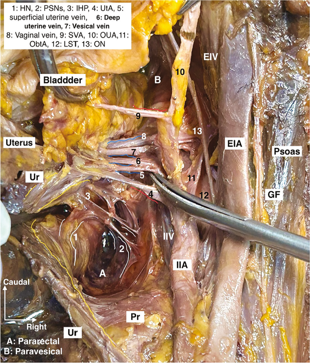
EIA: External iliac artery, EIV: External iliac vein, IIA: Internal iliac artery, IIV: Internal iliac vein, GF: Genitofemoral nerve, HN: Hypogastric nerve, IHP: Inferior hypogastric plexus, LST: Lumbosacral trunk, ObtA: Obturator artery, ON: Obturator nerve, OUA: Obliterated umbilical artery, Pr: Promontory, PSNs: Pelvic splanchnic nerves, SVA: Superior vesical artery, Ur: Ureter, UtA: Uterine artery
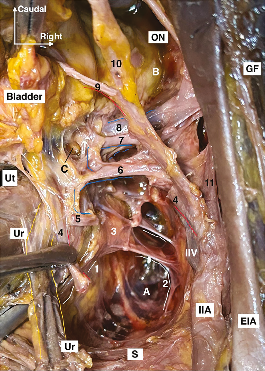
A: Pararectal space, B: Paravesical space, C: Paravaginal space, EIA: External iliac artery, IIA: Internal iliac artery, IIV: Internal iliac vein, GF: Genitofemoral nerve, ON: Obturator nerve, S: Sacrum, Ur: Ureter, Ut: Uterus, 1: Hypogastric nerve, 2: Pelvic splanchnic nerves, 3: Inferior hypogastric plexus, 4: Uterine artery, 5: Superficial uterine vein, 6: Deep uterine vein, 7: Vesical vein, 8: Vaginal vein, 9: Superior vesical artery, 10: Obliterated umbilical artery, 11: Obturator artery
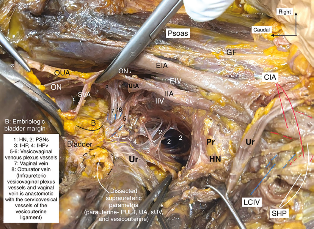
CIA: Common iliac artery, EIA: External iliac artery, EIV: External iliac vein, IIA: Internal iliac artery, IIV: Internal iliac vein, GF: Genitofemoral nerve, HN: Hypogastric nerve, LCIV: Left common iliac vein, ON: Obturator nerve, OUA: Obliterated umbilical artery, Pr: Promontory, PULT: Parauterine lymphatic tissue, SHP: Superior hypogastric plexus, sUV: Superficial uterine vein, SVA: Superior vesical artery, UA: Uterine artery, UtA: Uterine artery, Ur: Ureter, 1-HN: Hypogastric nerve, 2-PSNs: Pelvic splanchnic nerves, 3-IHP: Inferior hypogastric plexus, 4-IHP-v: Inferior hypogastric plexus vesical branches (bladder branches)
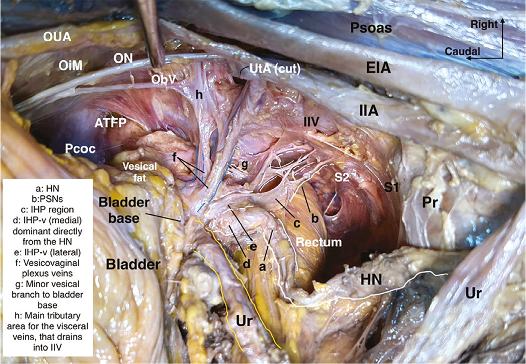
ATFP: Arcus tendineus fascia pelvis, EIA: External iliac artery, IIA: Internal iliac artery, IIV: Internal iliac vein, ObV: Obturator vein, OiM: Obturator internus muscle, ON: Obturator nerve, OUA: Obliterated umbilical artery, Pcoc: Pubococcygeus, Pr: Promontory, S: Sacrum, Ur: Ureter, UtA: Uterine artery, HN: Hypogastric nerve, a-HN: Hypogastric nerve, b-PSNs: Pelvic splanchnic nerves, c-IHP: Inferior hypogastric plexus, d-IHP-v: Inferior hypogastric plexus vesical branches (bladder branches)
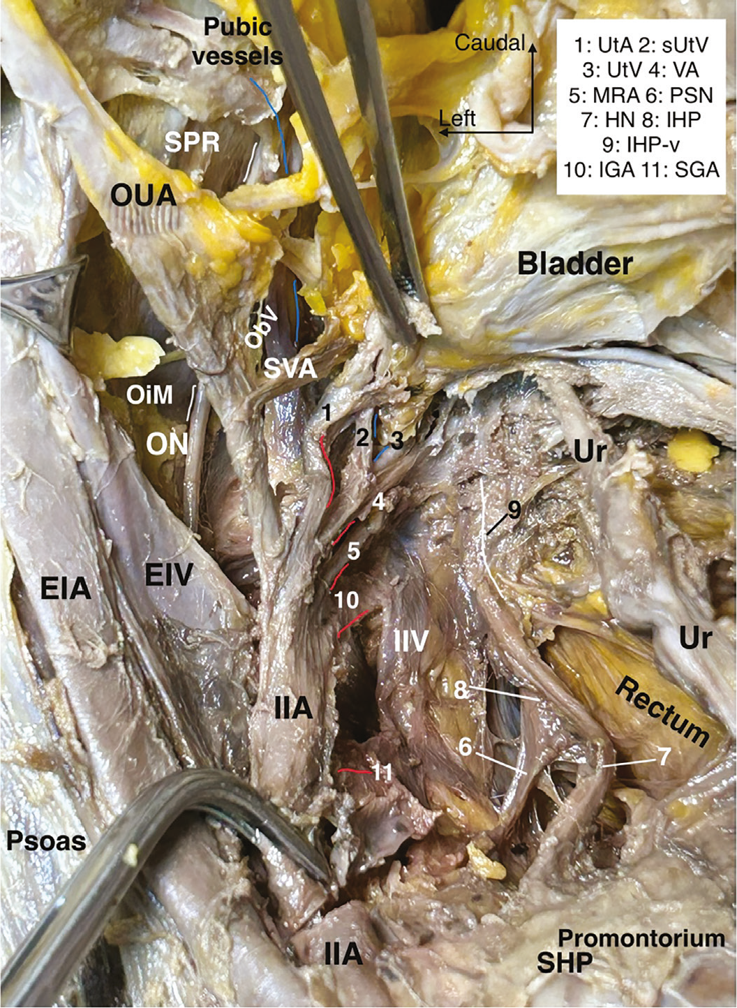
EIA: External iliac artery, EIV: External iliac vein, IIA: Internal iliac artery, IIV: Internal iliac vein, ON: Obturator nerve, OiM: Obturator internus muscle, ObV: Obturator vein, SPR: Superior pubic ramus, OUA: Obliterated umbilical artery, SVA: Superior vesical artery, SHP: Superior hypogastric plexus, Ur: Ureter, 1/UtA: Uterine artery, 2/sUtV: Superficial uterine vein, 3/UtV: Uterine vein, 4/VA: Vaginal artery, 5/MRA: Middle rectal artery, 6/PSN: Pelvic splanchnic nerves, 7/HN: Hypogastric nerve, 8/IHP: Inferior hypogastric plexus, v: Vesical branches, 10/IGA: Inferior gluteal artery, 11/SGA: Superior gluteal artery
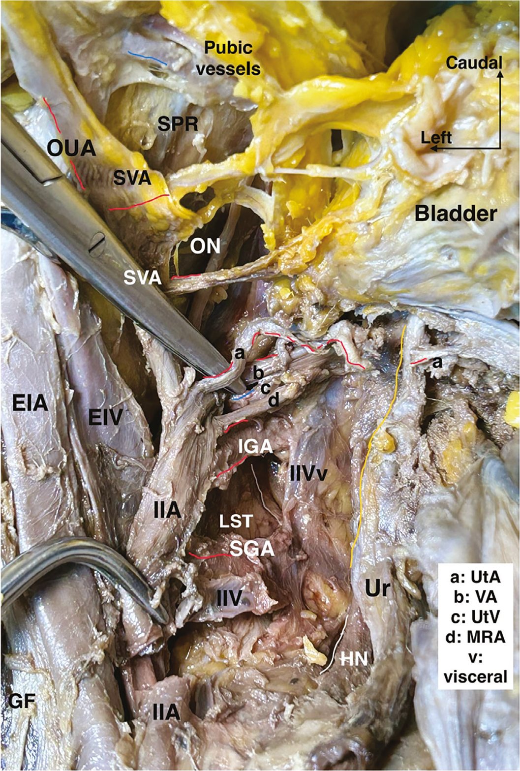
GF: Genitofemoral nerve, EIA: External iliac artery, EIV: External iliac vein, IIA: Internal iliac artery, IIV: Internal iliac vein, ON: Obturator nerve, SPR: Superior pubic ramus, OUA: Obliterated umbilical artery, SVA: Superior vesical artery, a/UtA: Uterine artery, b/VA: Vaginal artery, c/UtV: Uterine vein, d/MRA: Middle rectal artery, IGA: Inferior gluteal artery, SGA: Superior gluteal artery, LST: Lumbosacral trunk, HN: Hypogastric nerve, Ur: Ureter
The pararectal space serves as the gateway to preservation of pelvic autonomic nerves; correct development of this space distinguishes modern nerve-sparing oncologic surgery from purely radical resection. Mastery of the paravesical space transforms pelvic surgery from an organ-based dissection to a space-oriented surgery, reducing morbidity while maintaining radicality.
Prevesical Space (Retropubic Space of Retzius) Definition and Surgical Concept
The prevesical space, also known as the retropubic space of Retzius, is a midline, anterior, extraperitoneal, avascular compartment located between the posterior surface of the pubic bone and the anterior wall of the urinary bladder. It represents the most anterior avascular space of the pelvis and constitutes the primary surgical corridor of the anterior pelvic compartment. In pelvic surgery, the prevesical space is essential for anterior pelvic orientation, bladder mobilization, and access to the paravesical spaces.
Anatomical Boundaries ()
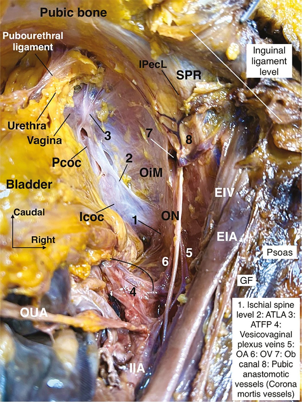
EIA: External iliac artery, EIV: External iliac vein, Icoc: Iliococcygeus, IIA: Internal iliac artery, IPecL: Pectineal ligament, GF: Genitofemoral nerve, OiM: Obturator internus muscle, ON: Obturator nerve, OUA: Obliterated umbilical artery, Pcoc: Pubococcygeus, SPR: Superior pubic ramus, ATLA: Arcus tendineus levator ani, ATFP: Arcus tendineus fascia pelvis, OA: Obturator artery, OV: Obturator vein, Ob: Obturator
• Anterior boundary:
Posterior surface of the pubic symphysis and pubic bone.
• Posterior boundary:
Anterior wall of the urinary bladder.
• Lateral boundaries:
Pubocervical ligaments,
OUA,
Transition to the bilateral paravesical spaces.
• Superior boundary:
Anterior parietal peritoneum, reflection over the bladder dome.
• Inferior boundary:
Pelvic floor,
Endopelvic fascia(corresponds to the en bloc structure of the visceral fascia of the bladder and pelvic parietal fascia over the levator ani, pubocervical fascia),
The inferolateral border is the tendinous arch of pelvic fascia,
These boundaries position the prevesical space as a central, anterior, avascular plane that is continuous laterally with the paravesical spaces.
Surgical Development (Step-by-Step)
1. Anterior peritoneal mobilization
The bladder peritoneum is gently mobilized caudally from the anterior abdominal wall at the posterior aspect of the rectus abdominis muscle. The urachus is visible, extending from the bladder apex to the umbilicus.
This exposes the extraperitoneal plane above the bladder dome.
2. Midline entry
Blunt dissection is initiated in the midline between the bladder and pubic symphysis,
Loose areolar tissue allows atraumatic entry, slightly lateral to the bladder neck, bilaterally.
3. Space expansion
Dissection proceeds caudally, along the lateral aspect of the urethra,
The bladder is gently displaced posteriorly.
4. Lateral extension
The space is widened bilaterally,
Continuity with the paravesical spaces is established,
Correct development creates a bloodless retropubic working space, enabling safe anterior pelvic surgery.
Contents and Key Structures
The prevesical space itself is avascular but contains and is closely related to critical structures:
• Vascular
Retropubic venous plexus (Santorini plexus), lateral to the urethra at the deep paravaginal compartment,
Small pubic veins.
• Fascial
Endopelvic fascia, pubocervical fascia,
Pubovesical ligaments.
• Visceral
Urinary bladder, posteriorly,
Because of the proximity of the retropubic venous plexus, meticulous blunt dissection is essential to avoid significant bleeding.
Surgical Applications ()
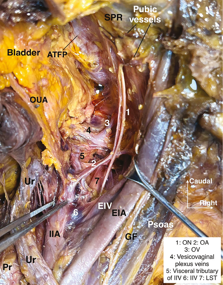
GF: Genitofemoral nerve, EIA: External iliac artery, EIV: External iliac vein, IIA: Internal iliac artery, IIV: Internal iliac vein, Ur: Ureter, Pr: Promontorium, OUA: Obliterated umbilical artery, ATFP: Arcus tendineus fascia pelvis, SPR: Superior pubic ramus, ON: Obturator nerve, OA: Obturator artery, OV: Obturator vein, LST: Lumbosacral trunk
The prevesical space has multiple important surgical applications:
• Radical hysterectomy
Enables anterior bladder mobilization,
Facilitates the separation of the bladder from the cervix and upper vagina in complex cases.
• Pelvic lymphadenectomy
Provides orientation for anterior pelvic anatomy before entering paravesical spaces in frozen pelvis cases.
• Hemorrhage control
Allows exposure and compression of the retropubic venous structures.
• Pelvic exenteration
Serves as the anterior dissection plane in anterior or total pelvic exenteration.
• Reconstructive and urogynecologic procedures
Provides access for retropubic sling placement (conceptually relevant for anatomical orientation), transvaginal tape procedure,
Pectineal ligament suspension (Cooper’s ligament, Burch procedure),
These applications highlight the prevesical space as a foundational anterior pelvic corridor rather than a simple anatomical cavity.
Key Surgical Insight
The prevesical space is the anatomical starting point of anterior pelvic surgery; gentle midline blunt dissection and respect for the retropubic venous plexus are essential to maintain a bloodless field.
Medial Psoas Space (Laterovascular Plane) Definition and Surgical Concept
The medial psoas space, also referred to as the laterovascular plane, is a retroperitoneal anatomical plane located medial to the psoas major muscle and lateral to the iliac vessels. It is not a classical “avascular space,” such as the paravesical or pararectal spaces, but rather a longitudinal dissection plane that serves as the lateral boundary for pelvic vascular and lymphatic dissection. This plane is fundamental for pelvic and paracervical lymphadenectomy, for safe exposure of the iliac vessels, the obturator region (including the obturator nerve) and the lumbosacral trunk, and for providing a stable anatomical reference along the pelvic sidewall.
Anatomical Boundaries ()
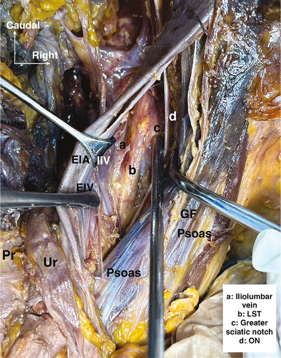
EIA: External iliac artery, EIV: External iliac vein, IIV: Internal iliac vein, GF: Genitofemoral nerve, Pr: Promontory, Ur: Ureter, LST: Lumbosacral trunk, ON: Obturator nerve
The medial psoas space is defined by consistent muscular, vascular, and fascial landmarks:
• Lateral boundary:
Psoas major muscle,
Psoas fascia.
• Medial boundary:
External iliac artery and vein,
Internal iliac vein,
Common iliac vessels (cranially).
• Anterior boundary:
Retroperitoneal fatty-lymphatic tissue.
• Posterior boundary:
Psoas major muscle body.
• Superior boundary:
Pelvic brim, linea terminalis,
Overlying parietal peritoneum.
• Inferior boundary:
Obturator fossa region,
These boundaries position the medial psoas space as the lateral limit of pelvic oncologic dissection and the entry plane into the obturator and sacral compartments.
Surgical Development (Step-by-Step)
1. Retroperitoneal entry
Achieved by incising the pelvic lateral parietal peritoneum:
• After transection of the round ligament, or,
• Between the round ligament and the infundibulopelvic ligament.
2. Identification of the psoas major
The psoas major muscle is visualized as the first constant structure,
Its medial border defines the lateral limit of safe pelvic dissection.
3. Exposure of the vascular structures
The external iliac artery and vein are identified medial to the psoas major muscle,
Retroperitoneal lymphatic tissue and external iliac vessels are gently mobilized medially (during this step, a psoas vessel branch can be noticed).
4. Development of the laterovascular plane
Blunt dissection proceeds along the medial surface of the psoas major muscle,
This creates a longitudinal plane separating the iliac vessels from the muscle.
5. Caudal extension
The plane is followed inferiorly toward the obturator fossa,
The obturator nerve becomes visible at the superficial caudal extent, where it emerges from the posteromedial part of the psoas muscle,
Medially, the internal iliac vein can be noticed,
Inferior to the obturator nerve, the lumboscaral trunk can be dissected, at the inferomedial part cranially, and at the inferolateral part caudally,
Between the obturator nerve and the lumbosacral trunk, the iliolumbar vessels may lie. they drain into the internal iliac vein, Correct development produces a clean, tension-free lateral boundary, enabling safe nodal dissection without vascular or neural injury. Furthermore, the laterovascular plane and the caudal region of the lumbosacral trunk are accessible through the medial approach (). During the surgical procedure, the internal iliac artery and vein, along with their associated vessels, may be sacrificed entirely or partially. Additionally, the inter-iliac region, situated between the external and internal iliac vessels, may be utilized.
Contents and Related Key Structures ()
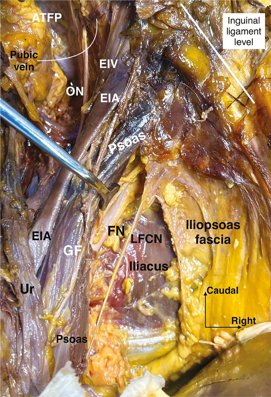
ATFP: Arcus tendineus fascia pelvis, EIA: External iliac artery, EIV: External iliac vein, FN: Femoral nerve, GF: Genitofemoral nerve, LFCN: Lateral femoral cutaneous nerve, ON: Obturator nerve, Ur: Ureter
The medial psoas space contains or exposes several critical structures:
• Neural
Genitofemoral nerve, running on the anterior surface of the psoas muscle, lateral to the external iliac artery,
Obturator nerve, caudally, at the obturator fossa,
Lumbosacral trunk, caudally, inferior to the obturator nerve, merges with the sacral 1-3 nerves to form the sciatic nerve at the infrapiriform part of the greater sciatic foramen.
• Vascular
External iliac artery and vein, medially,
Internal iliac vein, medially, inferior to the external iliac vein,
Femoral nerve is a relevant anatomical structure noticed after dissection of the iliopsoas fascia, between the psoas and iliacus muscles, located at the posterolateral aspect of the psoas major muscle.
• Urologic
Ureter, medial to the iliac vessels, crossing the pelvic brim, is a related adjacent visceral tissue.
• Lymphatic
External iliac lymph nodes,
Obturator lymphatic tissue, distally and caudally,
Paracervical lymph nodes between the obturator nerve and the lumbosacral trunk,
Deep common iliac lymph nodes,
Recognition of these structures is essential to maintain oncologic radicality while preserving nerve integrity.
Surgical Applications
The medial psoas space (laterovascular plane) has multiple key surgical roles:
• Pelvic lymphadenectomy
Defines the lateral limit of the external iliac and cranial obturator nodal dissection,
Guides entry into the obturator fossa, from the lateral perspective.
• Radical hysterectomy
Provides orientation for safe parametrial dissection,
Protects the genitofemoral and obturator nerves,
Provides paracervical lymphadenectomy,
Provides type D resection, laterally extended parametrectomy, and endopelvic resection.
• Ureteral identification
Facilitates a reliable localization of the ureter at the pelvic brim in complex cases.
• Hemorrhage control
Maintains a clear boundary between the lymphatic tissue and major vessels.
• Advanced pelvic surgery
Serves as the lateral reference plane when extending the dissection cranially.
• Neuropelviology
Important for sciatica-related nodules and masses.
Key Surgical Insight ()
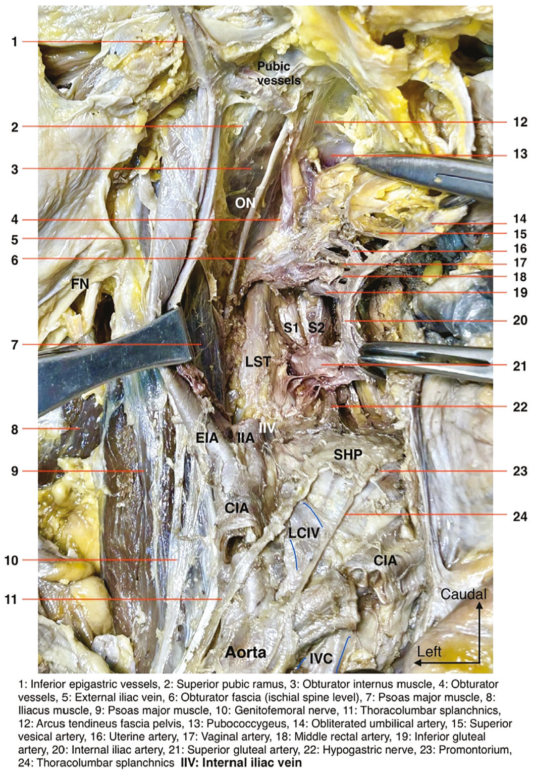
IVC: Inferior vena cava, CIA: Common iliac artery, LCIV: Left common iliac vein, SHP: Superior hypogastric plexus, EIA: External iliac artery, IIA: Internal iliac artery, IIV: Internal iliac vein, LST: Lumbosacral trunk, S: Sacral, ON: Obturator nerve, FN: Femoral nerve
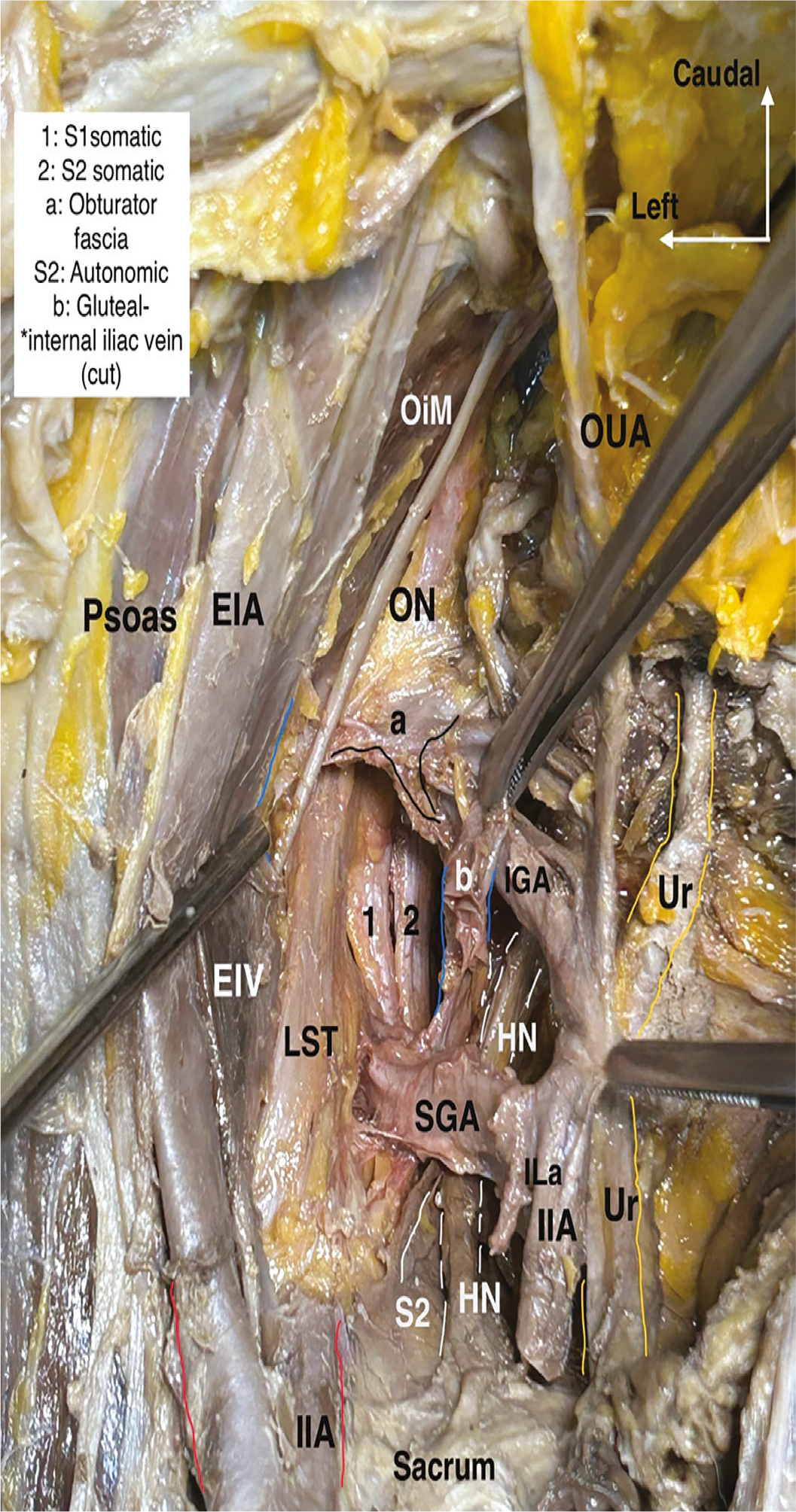
EIA: External iliac artery, EIV: External iliac vein, IIA: Internal iliac artery, OiM: Obturator internus muscle, ON: Obturator nerve, OUA: Obliterated umbilical artery, Ur: Ureter, S: Sacral, HN: Hypogastric nerve, ILa: Iliolumbar artery, SGA: Superior gluteal artery, IGA: Inferior gluteal artery, LST: Lumbosacral trunk
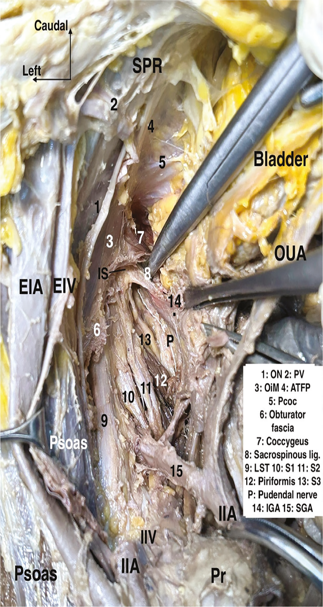
EIA: External iliac artery, EIV: External iliac vein, IIA: Internal iliac artery, IIV: Internal iliac vein, IS: Ischial spine, OUA: Obliterated umbilical artery, P: Pudendal, Pr: Promontory, SPR: Superior pubic ramus, 1-ON: Obturator nerve, 2-PV: Pubic vessels, 3-OiM: Obturator internus muscle, 4-ATFP: Arcus tendineus fascia pelvis, 5-Pcoc: Pubococcygeus, 6: Obturator fascia, 7: Coccygeus, 8: Sacrospinous ligament, 9-LST: Lumbosacral trunk, 10-S1: Sacral 1, 11-S2: Sacral 2, 12: Piriformis, 13-S3: Sacral 3, 14-IGA: Inferior gluteal artery, 15-SGA: Superior gluteal artery, lig: Ligament
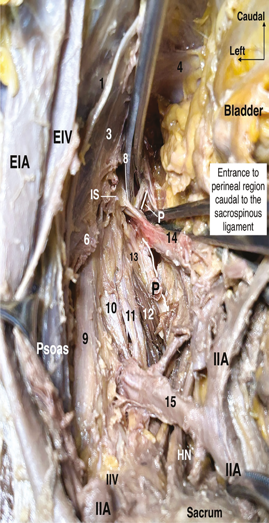
EIA: External iliac artery, EIV: External iliac vein, IIA: Internal iliac artery, IIV: Internal iliac vein, HN: Hypogastric nerve, IS: Ischial spine, P: Pudendal, 1-ON: Obturator nerve, 3-OiM: Obturator internus muscle, 4-ATFP: Arcus tendineus fascia pelvis, 6: Obturator fascia, 8: Sacrospinous ligament, 9-LST: Lumbosacral trunk, 10: Sacral 1, 11: Sacral 2, 12: Piriformis, 13: Sacral 3, 14-IGA: Inferior gluteal artery, 15-SGA: Superior gluteal artery
The medial psoas space is the lateral guide for pelvic oncologic surgery; respecting this plane ensures vascular safety, nerve preservation, and oncologic precision.
What does This Study Add to the Literature, and Limitations
The pelvic anatomy is a complex, three-dimensional structure, which complicates its understanding. This complexity may lead to complications during pelvic surgeries. Although numerous anatomical reports have been published, this study aims to enhance understanding of surgical anatomy through a systematic, layer-by-layer dissection progressing from superficial to deep structures. It includes a comprehensive evaluation of pelvic avascular spaces, namely the presacral, pararectal, paravesical, and prevesical. In reviewing surgical anatomy, this study emphasizes the critical anatomical structures and associated surgical maneuvers, employing cadaveric dissection to detail anatomical landmarks. The primary limitation of this study is that it assesses surgical practice solely through photographs of cadaveric dissection.
Nevertheless, this represents the primary strength of the study, which was originally designed to delineate the anatomical points using to observe details not identifiable during live surgery.
Conclusion
The pelvic anatomy exhibits consistency. All the retroperitoneal compartments—including the presacral, pararectal, paravesical, and prevesical spaces—are interconnected. The laterovascular plane, also known as the medial psoas space, is part of the lateral pelvic spaces (pararectal and paravesical). Key anatomical landmarks for identifying retroperitoneal structures are the sacral promontory posteriorly (representing the anterior edge of the first sacral vertebra at the level of the pelvic brim), the pubic symphysis or the posterior surface of the body of the pubis anteriorly (serving as the superior midline component of the pubic bone), and the psoas major muscle laterally. Improved knowledge of retroperitoneal surgical anatomy plays a crucial role in safe pelvic surgery.
Footnotes
Authorship Contributions
Surgical and Medical Practices: İ.S., Ö.Ö., N.K., M.A.G., M.D., Concept: İ.S., Ö.Ö., N.K., M.A.G., M.D., Design: İ.S., Ö.Ö., N.K., M.A.G., M.D., Data Collection or Processing: İ.S., Ö.Ö., N.K., M.A.G., M.D., Analysis or Interpretation: İ.S., Ö.Ö., N.K., M.A.G., M.D., Literature Search: İ.S., Ö.Ö., N.K., Writing: İ.S.
Conflict of Interest: No conflict of interest was declared by the authors.
Financial Disclosure: The authors declared that this study received no financial support.
References
Selçuk İ, Ersak B, Tatar İ, Güngör T, Huri E. Basic clinical retroperitoneal anatomy for pelvic surgeons. Turk J Obstet Gynecol. 2018;15:259-69.