
Abstract
Endometriosis is a common condition among women and can cause complications such as abdominal pain, dysmenorrhea, and infertility. One of the potential causes of this disease is autoimmunity. However, evidence regarding the role of autoimmunity is conflicting and inconclusive. The aim of this study was to investigate whether autoantibodies, a sign of autoimmunity, are present in people suffering from endometriosis. Relevant studies up to April 14, 2023 were identified by systematically searching Scopus, PubMed, Web of Science, Embase, and Google Scholar. This meta-analysis includes all qualified case-control studies of human populations that analyzed the association between serum autoantibodies and endometriosis. The odd ratios and 95% confidence intervals were calculated. In addition, heterogeneity and publication bias were examined, and subgroup analyses were performed based on region and target antigens. Forty-one studies were included, comparing 2,825 endometriosis patients with 4,158 healthy controls. The meta-analysis findings indicated a significant association between the presence of autoantibodies in the serum and an increased susceptibility to endometriosis (odds ratio: 4.242, confidence interval 95%: 3.824-4.706, p<0.001). In addition, there was a significant correlation between the presence of endometriosis and serum levels of anti-nuclear antibodies, B2 glycoprotein 1, CA125, carbonic anhydrase 1, cardiolipin, endometrial, laminin-1, smooth muscle, and syntaxin autoantibodies. Upon further analysis, it was found that the serum levels of these autoantibodies were higher in patients with endometriosis from North America than in those from other regions (p=0.001). The study revealed a significant correlation between serum autoantibodies and susceptibility to endometriosis, highlighting autoimmunity as a potential cause.
Keywords: Autoantibody, endometriosis, meta-analysis, serum
Introduction
Endometriosis is a prevalent medical condition characterized by endometrial tissue in extrauterine locations(). This misplaced tissue responds to hormones and can cause internal bleeding, inflammation, and fibrosis(). A definitive diagnosis of endometriosis is performed after biopsy by surgery; therefore, its exact prevalence is uncertain(). However, it is estimated to affect 10-15% of women of reproductive age(). People affected by this disease experience symptoms such as pelvic pain, dysmenorrhea, and infertility().
The exact etiology of this endometriosis remains unknown. However, previous research has shown that both genetic and environmental factors contribute to its development. Epigenetic abnormalities, anomalous estrogen production, retrograde menstruation, autoimmune responses, and allergic reactions are potential etiological factors(, ).
Several studies have been conducted on the role of autoimmune responses as a potential etiological factor in endometriosis. They have demonstrated that endometriosis is correlated with persistent regional inflammation and autoantibodies. Their results showed that women with endometriosis display immune system abnormalities similar to those seen in autoimmune diseases(, ). This includes disrupted immune surveillance, abnormal T and B-cell functions, heightened humoral immune response with increased autoantibodies in the serum, and inflammatory tissue damage(, ).
Although several studies have explored the connection between the presence of autoantibodies in the serum and the likelihood of developing endometriosis, the results have been inconsistent. The aim of this study was to analyze all relevant research and perform a meta-analysis to investigate this association.
Materials and Methods
Eligibility Criteria, Information Sources, and Search Strategy
This meta-analysis followed the Preferred Reporting Items for Systematic Reviews and Meta-analysis guidelines(). Various databases, including Scopus, PubMed, Web of Science, EMBASE, and 30 pages of Google Scholar, were searched to identify relevant studies. The search was not restricted by language and covered the period from April 2023. The references of the included studies were also hand-searched. The search strategies consisted of the terms “endometriosis”, “autoantibodies”, “serum” and their related synonyms.
Women diagnosed with endometriosis based on standard criteria confirmed by laparoscopic sampling and/or histological examination will participate in this review. This study will compare endometriosis patients with healthy women of the same age group.
Study Selection
This meta-analysis included all available case-control studies that met the following criteria: Analysis of the association between autoantibodies and endometriosis patients provides necessary information for a meta-analysis.
The exclusion criteria were as follows: Letters, editorials, abstracts, conference abstracts, or publications lacking adequate information; studies that used women with diseases other than endometriosis, particularly autoimmune diseases; and studies that explored the presence of autoantibodies in bodily fluids or tissues other than serum.
Data Extraction
Two reviewers independently extracted the pertinent data from the selected studies. If necessary, the investigators resolved the differences through discussion and consultation with a third-party. The information extracted was entered into an Excel form, including the last name of the first author, date and location of the study, number of participants in both the case and control groups, mean age of participants, method of diagnosing endometriosis, subclass of autoantibodies, sample type, method of evaluating autoantibodies, and the number or mean value and standard deviation of autoantibody-positive cases and controls.
Assessment of the Risk of Bias
The articles’ quality was evaluated by two reviewers using the Newcastle-Ottawa Quality Assessment scale (NOS)(), and any discrepancies were resolved by a third reviewer. Articles that achieved a NOS score of 7 or above were classified as high quality, whereas those with scores between 5 and 7 were moderate quality.
Data Synthesis
All data were statistically analyzed using Comprehensive meta-analysis software version 3.0 (Biostat, USA). To determine the odds ratios (ORS) and their corresponding 95% confidence intervals (CIS), the statistical analysis used the groups’ sample size, mean serum autoantibody level, and standard deviation for both patients diagnosed with endometriosis and the healthy control group. Some studies presented continuous variables as median and quartile, which were converted to mean and standard deviation using the formula in the Cochrane Handbook(, ). The heterogeneity of the studies was assessed using Cochrane Q and I2 statistics. The random-effects model was used to estimate the outcome data in cases where the Cochrane Q p-value was less than 0.1 and the I2 value exceeded 50%, indicating the presence of statistical heterogeneity. Conversely, a fixed-effects model was employed in other instances. Subgroup analysis was performed to evaluate the impact of confounding variables on the outcomes of the meta-analysis. A sensitivity analysis was also conducted, in which each study was systematically excluded to assess the stability of the obtained results.
Results
Study Selection
After conducting a comprehensive search of the available resources, 1,053 studies were obtained. We then removed any duplicate sources and examined the titles and abstracts of 663 studies. Of these, 71 studies were selected for full-text review. Finally, after applying the inclusion and exclusion criteria, 41 studies were included in our meta-analysis. The process of literature screening and its outcomes are presented in . The studies included in the analysis were assigned ratings ranging from zero to nine based on the Ottawa-Newcastle scale for case-control studies. As shown in , thirty-three studies were assessed as being of high quality, whereas eight studies were evaluated as being of moderate quality.
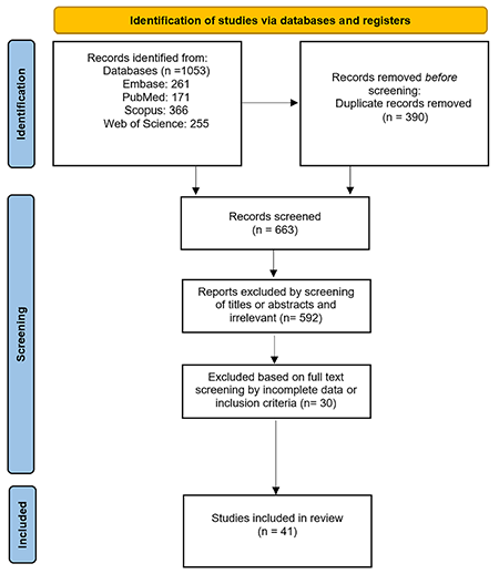
Table 1. Basic characteristics of the included studies
First author | Year | Country | Ethnicity | Endometriosis diagnosis protocol | Mean age or age range case | Mean age or age range control | NOS score |
|---|---|---|---|---|---|---|---|
Mathur et al.(44) | 1982 | USA | North American | Laparoscoy and biopsy or laparotomy | 26-37 | 26-37 | 6 |
Taylor et al.(15) | 1991 | UK | European | Laparoscoy and biopsy or laparotomy | 33.5 | 31.6 | 6 |
Gorai et al.(33) | 1993 | Japan | Asian | Laparoscopy or laparotomy | 21-46 | 20-46 | 7 |
Fernández-Shaw et al.(16) | 1993 | UK | European | Laparoscopy | - | - | 7 |
Kiechle et al.(45) | 1994 | USA | North American | Laparoscoy | 34.8 | 32.6 | 7 |
Kim et al.(34) | 1995 | Korea | Asian | Laparoscopy or laparotomy | 30-40 | - | 6 |
Aoki et al.(46) | 1995 | USA | North American | Laparoscopy | 33 | 33 | 7 |
Nip et al.(17) | 1995 | UK | European | Laparoscopy | 34 | 34 | 8 |
Odukoya et al.(18) | 1995 | UK | European | Laparoscopy and histology | 33 | 33 | 7 |
Pillai et al.(50) | 1996 | USA | North American | Laparoscopy | - | 25-35 | 6 |
Hatayama et al.(35) | 1996 | Japan | Asian | Laparoscopy or laparotomy | 35.3 | 35.3 | 6 |
D’Cruz et al.(47) | 1996 | USA | North American | Laparoscopy | <45 | <45 | 7 |
Fernández-Shaw et al.(19) | 1996 | UK | European | Laparoscopy | - | - | 7 |
Kim et al.(36) | 1997 | Korea | Asian | Laparoscopy | 32 | 32 | 7 |
Kaider et al.(48) | 1999 | USA | North American | Laparoscopy | - | - | 8 |
Shanti et al.(51) | 1999 | USA | North American | Histologically | 18-45 | - | 7 |
Szczepañska et al.(25) | 2001 | Poland | European | Laparoscopy | 29.0 | - | 7 |
Reimand et al.(26) | 2001 | Estonia | European | Laparoscopy and histology | 27.5 | 31.0 | 8 |
Ulcová-Gallová et al.(32) | 2002 | Czech Republic | European | Laparoscopy | 35.6 | 35.6 | 6 |
Pasoto et al.(52) | 2002 | Brazil | South American | Laparoscopy | 18-40 | 18-40 | 7 |
Inagaki et al.(43) | 2003 | Israel | Asian | Laparoscopy and laparotomy | 33.7 | 29.6 | 7 |
Pasoto et al.(53) | 2005 | Brazil | South American | Laparoscopy or laparotomy and histological | 18-40 | 18-40 | 7 |
Haller et al.(27) | 2006 | Estonia | European | Laparoscopy | 33.0±5.5 | - | 7 |
Randall et al.(49) | 2007 | USA | North American | Laparoscopy | 31.8±6.5 | 31.8±6.5 | 7 |
Haller et al.(28) | 2007 | Estonia | European | Laparoscopy | 34.0±4.8 | 44.9±10.7 | 8 |
Caccavo et al.(20) | 2007 | Italy | European | Laparoscopy | 34.4±4.2 | 33.3±3.1 | 7 |
Aguiar et al.(54) | 2009 | Brazil | South American | laparoscopy and histology | 29.2±5.6 | 27.9±4.5 | 8 |
Nabeta et al.(37) | 2009 | Japan | Asian | Laparoscopy | 34.7±7.6 | 35.0±3.7 | 6 |
Nabeta et al.(38) | 2010 | Japan | Asian | laparosccopy or laparotomy | 35.2±8.0 | 34.2±9.3 | 7 |
Inagaki et al.(41) | 2011 | Japan | Asian | Laparoscopy or laparotomy and histology | 31.9 | 29.6 | 9 |
Caccavo et al.(21) | 2011 | Italy | European | laparoscopy and histology | 34.8±3.7 | - | 7 |
Nabeta et al.(39) | 2011 | Japan | Asian | Laparoscopy or laparotomy | 35.2±8.0 | 34.2±9.3 | 7 |
Gajbhiye et al.(42) | 2012 | India | Asian | Laparoscopy and histology | 31.2±4.9 | 28.9±5.3 | 8 |
Ozhan et al.(22) | 2014 | Turkey | European | Laparoscopy or laparotomy | 32.33±7.01 | 34.20±6.88 | 7 |
Gajbhiye et al.(40) | 2017 | India | Asian | Laparoscopy and histology | 32.6±6.3 | 32.13±6.03 | 8 |
Yu-Rice et al.(29) | 2017 | USA & Germany | North American and European | - | 28-70 | 18-65 | 7 |
Ek et al.(23) | 2019 | Sweden | European | Laparoscopy | 38.0 | 42.5 | 7 |
Toullec et al.(30) | 2020 | French | European | Histologically | 33.00±5.365 | - | 7 |
Artymuk et al.(31) | 2021 | Russia | European | Laparoscopy and histology | 31.6±4.8 | 31.8±6.5 | 6 |
Svensson et al.(24) | 2022 | Sweden | European | Laparoscopy | 32.0-43.0 | 33-53 | 7 |
Vilas Boas et al.(55) | 2022 | Brazil | South American | Laparoscopy and histology | 37.2±7.1 | 37.8±5.6 | 8 |
NOS: Newcastle-Ottawa Quality Assessment scale
Study Characteristics
In line with the research methodology, we included a total of forty-one eligible case-control studies to investigate the correlation between the presence of autoantibodies and the risk of endometriosis. Eighteen studies were conducted in Europe(-), eleven in Asia(-), nine in North America(, -), and four in South America(-). The basic characteristics of the included studies are listed in and . shows the number of studies investigating each autoantibody.
Table 2. The fundamental details of the selected studies about the evaluation of autoantibodies
First author | Year | Case number | Control number | Ig type | Sample type | Method | The investigated autoantibody |
|---|---|---|---|---|---|---|---|
Mathur et al.(44) | 1982 | 13 | 15 | IgG | Serum | Passive haemagglutination | Endometrial Ovary Granulosa Theca |
Taylor et al.(15) | 1991 | 71 | 109 | IgG, IgM and IgA | Serum | IF, WB, Counterimmunoelectrophoresis and Double immunodiffusion | Cardiolipin ANA Lupus anti-coagulant Smooth muscle RO (SS-A) La (SS-B) |
Gorai et al.(33) | 1993 | 18 | 27 | - | Serum | WB | Endometrial |
Fernández-Shaw et al.(16) | 1993 | 13 | 7 | IgG | Serum | IHC | Endometrial Endothelial |
Kiechle et al.(45) | 1994 | 23 | 17 | IgG | Serum | WB | Carbonic anhydrase I |
Kim et al.(34) | 1995 | 33 | 20 | IgG | Serum | IHC and WB | Endometrial |
Aoki et al.(46) | 1995 | 64 | 97 | IgG | Serum | ELISA | Cardiolipin B2 Glycoprotein 1 Antiphospholipid antigens (PA, PG, PE, PI, PS) |
Nip et al.(17) | 1995 | 20 | 20 | IgG and IgM | Serum | ELISA and IF | Cardiolipin ANA Smooth muscle |
Odukoya et al.(18) | 1995 | 55 | 43 | IgG | Serum | ELISA | Endometrial Ovary Thyroid Skeletal muscle |
Pillai et al.(50) | 1996 | 46 | 18 | IgG | Serum | Passive hemagglutination | Endometrial Transferrin Alpha 2-Heremans Schmidt (HS) Glycoprotein |
Hatayama et al.(35) | 1996 | 20 | 20 | IgG | Serum | ELISA | Endometrial |
D’Cruz et al.(47) | 1996 | 319 | 100 | IgG | Serum | ELISA | Carbonic anhydrase I and II |
Fernandéz-Shaw et al.(19) | 1996 | 51 | 23 | IgG | Serum | ELISA | Endometrial |
Kim et al.(36) | 1997 | 42 | 87 | IgG | Serum | ELISA | Cardiolipin ANA Lupus anti-coagulant |
Kaider et al.(48) | 1999 | 23 | 105 | IgG, IgM and IgA | Serum | ELISA | Antiphospholipid antigens (PL) ANA Thyroid |
Shanti et al.(51) | 1999 | 40 | 16 | IgG | Serum | ELISA | Malondialdehyde-modified Oxidized low-density lipoprotein Lipid peroxide-modified |
Szczepañska et al.(25) | 2001 | 50 | 20 | IgG | Serum | ELISA | Zona pellucida |
Reimand et al.(26) | 2001 | 38 | 392 | IgG | Serum | IF and ELISA | B2 Glycoprotein 1 Carbonic anhydrase I ANA Smooth muscle Parietal cell Thyroid microsomal Mitochondrial |
Ulcová-Gallová et al.(32) | 2002 | 323 | 101 | IgG | Serum | ELISA | Cardiolipin B2 Glycoprotein 1 Antiphospholipid antigens (PA, PG, PE, PI, PS) Zona pellucida |
Pasoto et al.(52) | 2002 | 39 | 18 | - | Serum | IF | ANA RO/LA RNP/Sm dsNDA |
Inagaki et al.(43) | 2003 | 42 | 39 | IgG | Serum | ELISA | Laminin-1 |
Pasoto et al.(53) | 2005 | 45 | 21 | IgG and IgM | Serum | IF and ELISA | Cardiolipin Carbonic anhydrase I ANA RO (SS-A) dsDNA Histon Smooth muscle U1RNP |
Haller et al.(27) | 2006 | 12 | 56 | IgG | Serum | IF and ELISA | ANA Ovary Smooth muscle Parietal cell Thyroid microsomal Mitochondrial B2 Glycoprotein I Cardiolipin |
Randall et al.(49) | 2007 | 278 | 249 | IgG | Serum | IF | Endometrial |
Haller et al.(28) | 2007 | 12 | 85 | IgG, IgA and IgM | Serum | ELISA and IF and ImmunoCAP technology | FSH ANA Ovary Smooth muscle Parietal cell Thyroid microsomal Mitochondrial B2 Glycoprotein I Cardiolipin |
Caccavo et al.(20) | 2007 | 18 | 16 | IgG and IgM | Serum | ELISA | Cardiolipin |
Aguiar et al.(54) | 2009 | 120 | 1500 | IgA | Serum | ELISA and IF | Human tissue transglutaminase Endomysium |
Nabeta et al.(37) | 2009 | 65 | 70 | IgG | Serum | ELISA | CA125 Enolase |
Nabeta et al.(38) | 2010 | 69 | 82 | IgG | Serum | ELISA | CA125 PDIK1L |
Inagaki et al.(41) | 2011 | 45 | 39 | IgG | Serum | ELISA | Laminin-1 |
Caccavo et al.(21) | 2011 | 35 | 50 | IgG | Serum | ELISA | Laminin-1 |
Nabeta et al.(39) | 2011 | 69 | 82 | IgG | Serum | ELISA | Syntaxin |
Gajbhiye et al.(42) | 2012 | 50 | 27 | IgG | Serum | WB and ELISA | Tropomyosin 3 Stomatin-like protein 2 Tropomodulin 3 |
Ozhan et al.(22) | 2014 | 60 | 20 | - | Serum | ELISA | Laminin-1 Endometrial CA125 Syntaxin PDIK1L Enolase |
Gajbhiye et al.(40) | 2017 | 133 | 104 | - | Serum | ELISA | CA125 CA19-9 Syntaxin PDIK1L Enolase Tropomyosin 3a Tropomyosin 3b Tropomyosin 3c Tropomyosin 3d Stomatin-like protein 2a Stomatin-like protein 2b Stomatin-like protein 2c Tropomodulin 3a Tropomodulin 3b Tropomodulin 3c Tropomodulin 3d |
Yu-Rice et al.(29) | 2017 | 18 | 30 | IgG | Serum | ELISA | Selenium binding protein 1 |
Ek et al.(23) | 2019 | 100 | 100 | IgG | Serum | ELISA | LH LH receptor GnRH1 GnRH1 receptor Matrix metalloproteinase-9 Tenascin-C |
Toullec et al.(30) | 2020 | 106 | 92 | IgG | Serum | ELISA | Granulocyte-macrophage colony stimulating factor |
Artymuk et al.(31) | 2021 | 100 | 100 | IgG | Serum | ELISA | Estrogen Progesterone |
Svensson et al.(24) | 2022 | 53 | 50 | IgG | Serum | ELISA | LH LH receptor FSH FSH receptor TSH TSH receptor HCG |
Vilas Boas et al.(55) | 2022 | 94 | 91 | IgG | Serum | IF and ELISA | ANA ENA dsDNA |
Ig: Immunoglobulin, IF: Immunofluorescence, WB: Western-Blot, ELISA: Enzyme-Linked Immunosorbent Assay, IHC: Immunohistochemistry, FSH: Follicle-stimulating hormone,
LH: Luteinizing hormone, PDIK1L: PDLIM1 interacting kinase 1 like, ANA: Anti-nuclear antibodies, TSH: Thyroid-stimulating hormone, HCG: Human chorionic gonadotropin,
CA125: Carcinoembryonic antigen 125, CA19-9: Carcinoembryonic antigen 19-9, PA: Antiphospholipid antigen PA, PE: Antiphospholipid antigen PE, PG: Antiphospholipid antigen PG, PI: Antiphospholipid antigen PI, PL: Antiphospholipid antigen PL, PS: Antiphospholipid antigen PS, RO: A kind of extractable nuclear antigens (RO/SSA)
Table 3. The findings related to the analysis of heterogeneity and publication bias of the conducted studies
Variables (autoantibodies) | Number of included studies | I2 (%) | Q-test’s p-value | P-value of Begg’s test | P-value of Egger’s test |
|---|---|---|---|---|---|
All of the autoantibodies | 41 | 84.47 | <0.001 | 0.130 | 0.05 |
Anti-ANA | 8 | 0.00 | 0.859 | 0.804 | 0.406 |
Anti-B2 glycoprotein 1 | 3 | 0.00 | 0.578 | 0.601 | 0.169 |
Anti-CA125 | 4 | 75.25 | 0.007 | 0.174 | 0.415 |
Anti-carbonic anhydrase I | 4 | 36.39 | 0.194 | 1.000 | 0.663 |
Anti-cardiolipin | 7 | 1.51 | 0.413 | 0.880 | 0.746 |
Anti-endometrial | 9 | 93.07 | <0.001 | 0.251 | 0.788 |
Anti-enolase | 3 | 73.63 | 0.023 | 0.601 | 0.394 |
Anti-laminin-1 | 4 | 23.43 | 0.270 | 0.308 | 0.036 |
Anti-PDIK1L | 3 | 92.49 | <0.001 | 0.296 | 0.125 |
Anti-smooth muscle | 3 | 0.00 | 0.620 | 0.601 | 0.652 |
Anti-syntaxin | 3 | 79.70 | 0.007 | 0.601 | 0.940 |
ANA: Anti-nuclear antibodies, CA125: Carcinoembryonic antigen 125, PDIK1L: PDLIM1 interacting kinase 1 like
Risk of Bias of the Included Studies
Funnel plots, Begg’s rank correlation, and Egger’s regression tests were used to examine the existence of publication bias. and present the outcomes of publication bias. Based on the results of statistical tests and asymmetry analysis of funnel plots, there is a possible publication bias in the studies that investigated pooled and anti-laminin-1 autoantibodies (as shown in ). However, in other autoantibody studies, although a few exhibits slight visual asymmetry, statistical tests do not indicate any significant publication bias. The funnel plot diagrams, which show the likelihood of publication bias in studies on autoantibodies, were modified using the trim and fill test. The modifications did not result in any significant changes in studies that investigated the total and anti-laminin-1 autoantibodies (data not shown).
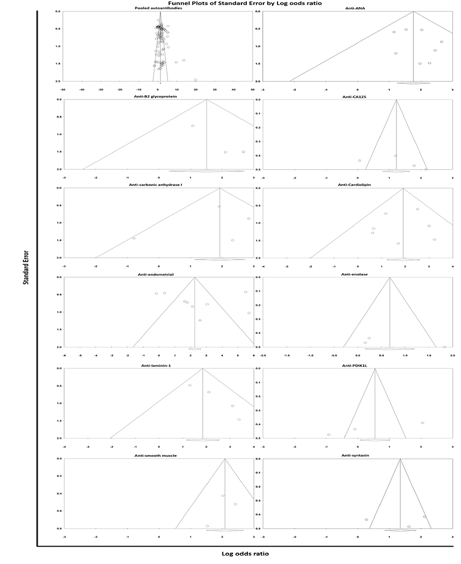
ANA: Anti-nuclear antibodies, CA125: Carcinoembryonic antigen 125, PDIK1L: PDLIM1 interacting kinase 1 like
Synthesis of the Results
The findings of the meta-analysis indicate a substantial correlation between the existence of autoantibodies and susceptibility to endometriosis (OR: 4.242, CI 95%: 3.824-4.706, p<0.001) (). Furthermore, individuals with endometriosis exhibited significantly higher levels of anti-nuclear antibodies (anti-ANA) (OR: 5.862, CI 95%: 3.454-9.950, p<0.001), B2 glycoprotein 1 (OR: 4.542, CI 95%: 1.360-15.175, p=0.014), CA125 (OR: 3.602, CI 95%: 1.485-8.733, p=0.005), carbonic anhydrase 1 (OR: 6.860, CI 95%: 3.043-15.468, p<0.001), cardiolipin (OR: 6.806, CI 95%: 3.369-13.749, p<0.001), endometrial (OR: 11.793, CI 95%: 2.382-58.383, p=0.002), laminin-1 (OR: 6.340, CI 95%: 3.151-12.757, p<0.001), smooth muscle (OR: 8.041, CI 95%: 4.442-14.557, p<0.001), and syntaxin (OR: 3.815, CI 95%: 1.249-11.649, p=0.019) autoantibodies than healthy controls. Statistical analysis found no significant relationship between autoantibodies against enolase (OR: 2.067, CI 95%: 0.739-5.782, p=0.167) and PDIK1L (PDLIM1 interacting kinase 1 like) (OR: 1.434, CI 95%: 0.241-8.528, p=0.692) and susceptibility to endometriosis.
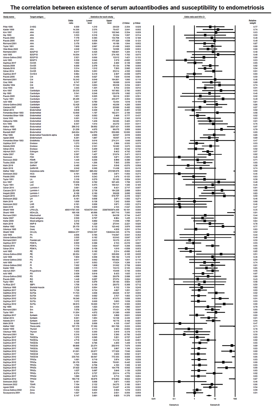
2-HSG: 2-Heremans Schmidt Glycoprotein, ANA: Anti-nuclear antibodies, B2GP1D: B2 glycoprotein 1 dependent, B2GP1I: B2 glycoprotein 1 independent, CA125: Carcinoembryonic antigen 125, CA19-9: Carcinoembryonic antigen 19-9, CAI, Carbonic anhydrase 1, CAII: Carbonic anhydrase II, FSH: Follicle-stimulating hormone, GM-CSF: Granulocyte-macrophage colony-stimulating factor, GnRH1: Gonadotropin releasing hormone 1, GnRH1R: Gonadotropin releasing hormone 1 receptor, HCG: Human chorionic gonadotropin, LA: One kind of neutrophile antigens (LA/SSB), LAC: Lupus anti-coagulant, LH: Luteinizing hormone, LHR: Luteinizing hormone receptor, LPM-LDL: Lipid peroxide-modified LDL, DMA-LDL: Malondialdehyde-modified LDL, MMP-9: Matrix metallopeptidase-9, OX-LDL: Oxidized low-density lipoprotein, PA: Antiphospholipid antigen PA, PDIK1L: PDLIM1 interacting kinase 1 like, PE: Antiphospholipid antigen PE, PG: Antiphospholipid antigen PG, PI: Antiphospholipid antigen PI, PL: Antiphospholipid antigen PL, PS: Antiphospholipid antigen PS, RO: A kind of extractable nuclear antigens (RO/SSA), SBP1: Selenium binding protein 1, SLP2a,b,c: Stomatin-like protein 2a,b,c ,Sm: Smooth muscle, TMA:, TMOD3a,b,c,d: Tropomodulin 3a,b,c,d, TPM3a,b,c,d: Tropomyosin 3a,b,c,d, TSH: Thyroid-stimulating hormone, TSHR: Thyroid-stimulating hormone receptor, t-TGA: Thyroglobulin
The relevant forest plots are presented in . It is worth mentioning that the conducted studies(, , ) failed to detect anti-dsDNA autoantibodies in individuals suffering from endometriosis, indicating the absence of any correlation.
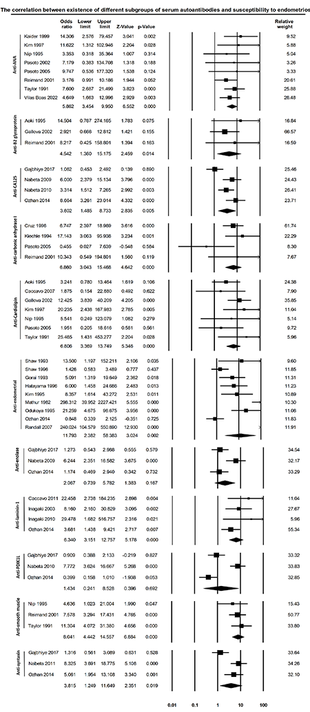
ANA: Anti-nuclear antibodies, CA125: Carcinoembryonic antigen 125, PDIK1L: PDLIM1 interacting kinase 1 like
Heterogeneity Test and Subgroup Analysis
The I2 test and Cochrane Q statistic showed heterogeneity among studies analyzing total, anti-CA125, anti-endometrial, anti-enolase, anti-PDIK1L, and anti-syntaxin autoantibodies. However, studies related to other autoantibodies showed no significant between-study heterogeneity, as shown in . To investigate the cause of heterogeneity, subgroup analysis was conducted based on the region. The findings revealed that patients with endometriosis from North America had notably higher levels of autoantibody titers than patients from other regions (p=0.001), and the region of living was identified as one of the sources of heterogeneity. In addition, subgroup analysis based on differences in target antigens of autoantibodies demonstrated this factor as a cause of heterogeneity in the study of pooled autoantibodies (p<0.001).
Sensitivity Analysis
Sensitivity analyses were conducted to evaluate the potential impact of a single study on the overall effect of autoantibodies in endometriosis. The sensitivity analyses indicated that upon exclusion of each study, the general conclusions remained substantially unchanged. These analyses consistently exhibited the robustness of the meta-analysis outcomes.
Discussion
Endometriosis is a significant health issue that affects women, but its exact cause remains unknown(). Several theories have been proposed to explain its origin, including autoimmunity(). However, studies investigating the development of autoantibodies in patients with autoimmune conditions have produced conflicting results. Therefore, the research collected and analyze the current body of information on this topic. The findings of our study indicate that the serum of individuals diagnosed with endometriosis has significantly higher levels of autoantibodies than that of healthy controls.
Studies have shown an increase in both the activity and quantity of B-cells in the serum of individuals with endometriosis(, ). The increase in B-cell activity leads to an elevated production of antibodies, which is consistent with the findings of the current study. Research has shown that immune cells, particularly B-cells, demonstrate an increase within lesions associated with endometriosis(). In addition, the secretion of cytokines responsible for activating B-cells, such as B lymphocyte-stimulators, also experiences an elevation within these specific regions(). These findings validate the results of our study.
Endometriosis relies on estrogen for its development() and estrogen has been identified as one of the potential mechanisms that increase the quantity and functionality of B-cells in individuals with endometriosis(). Research suggests that estrogen initiates a genetic program that alters the survival and activation of B-cells, leading to a shift in the naive immune system toward autoreactivity(). The exact cause of autoantibody formation in endometriosis is not well understood, but it is thought to be related to cellular damage and inflammation that occur in endometriosis-associated lesions. Abnormal exposure of self-antigens from damaged cells to the immune system triggers an autoimmune response, leading to the formation of autoantibodies that target cell-derived antigens().
This study suggests that differences in the target antigens of autoantibodies and continent of residence may contribute to the observed heterogeneity. The results of the subgroup analysis show that North American patients with endometriosis have higher levels of autoantibodies. Differences in genetic or environmental factors may explain this disparity. Additionally, according to the literature, autoimmune antibody manifestation varies among North American ethnic groups. Bruner et al.() discovered that African Americans with systemic lupus erythematosus exhibit significantly higher levels of anti-ANA autoantibody expression than other North American races. It is important to consider factors beyond ethnicity that can contribute to heterogeneity, such as sampling error, use of varied laboratory tests, body mass index, and age. It is crucial to study these factors in depth in future research efforts.
This meta-analysis is the most comprehensive study that systematically reviews and analyzes the relationship between autoantibodies and endometriosis, synthesizing over three decades of research. In addition, this study included a large sample size of 2,731 patients diagnosed with endometriosis and 4,067 healthy controls, potentially providing a definitive outcome with high accuracy and minimal bias for the general population.
It is important to note that there are limitations to this study. The laboratory data were measured in different centers using various methods and detection kits, which may have resulted in inconsistencies. Furthermore, the use of varying cutoff points across investigations could lead to inconsistencies in the outcome. In addition, the limited and insufficient data regarding the participants in the selected studies hindered our ability to examine other factors that may have an impact on heterogeneity, such as age and BMI. Therefore, future research should focus on exploring this aspect in more detail.
Conclusion
In conclusion, our meta-analysis revealed that patients diagnosed with endometriosis exhibit a greater prevalence of autoantibodies than healthy individuals. Based on the findings of this study, it is likely that autoimmune reactions are associated with the progression of endometriosis. However, further studies are required to determine the mechanism underlying autoantibody production in endometriosis. Future research should also investigate autoantibody levels during different phases of endometriosis.
Footnotes
Ethics
Authorship Contributions
Design: S.F., M.H.M., Data Collection or Processing: R.H.M., N.A., Analysis or Interpretation: S.F., R.H.M., N.A., M.H.M., Literature Search: M.H.M., Writing: S.F., R.H.M., N.A., M.H.M.
Conflict of Interest: No conflict of interest was declared by the authors.
Financial Disclosure: The authors declared that this study received no financial support.
References
Signorile PG, Viceconte R, Baldi A. New Insights in Pathogenesis of Endometriosis. Front Med (Lausanne) 2022;9:879015.
Arafah M, Rashid S, Akhtar M. Endometriosis: A Comprehensive Review. Adv Anat Pathol 2021;28:30-43.
Zondervan KT, Becker CM, Missmer SA. Endometriosis. N Engl J Med 2020;382:1244-56.
Parasar P, Ozcan P, Terry KL. Endometriosis: Epidemiology, Diagnosis and Clinical Management. Curr Obstet Gynecol Rep 2017;6:34-41.
Balun J, Dominick K, Cabral MD, Taubel D. Endometriosis in adolescents: a narrative review. Pediatr Med 2019;2:1-6.
Smolarz B, Szyłło K, Romanowicz H. Endometriosis: Epidemiology, Classification, Pathogenesis, Treatment and Genetics (Review of Literature). Int J Mol Sci 2021;22:10554.
Cousins FL, McKinnon BD, Mortlock S, Fitzgerald HC, Zhang C, Montgomery GW, et al. New concepts on the etiology of endometriosis. J Obstet Gynaecol Res 2023;49:1090-105.
Gajbhiye RK. Endometriosis and inflammatory immune responses: Indian experience. Am J Reprod Immunol 2023;89:e13590.
Nothnick WB. Treating endometriosis as an autoimmune disease. Fertil Steril 2001;76:223-31.
Abramiuk M, Grywalska E, Małkowska P, Sierawska O, Hrynkiewicz R, Niedźwiedzka-Rystwej P. The Role of the Immune System in the Development of Endometriosis. Cells 2022;11:2028.
Liberati A, Altman DG, Tetzlaff J, Mulrow C, Gotzsche PC, Loannidis JPA, et al. The PRISMA statement for reporting systematic reviews and meta-analyses of studies that evaluate health care interventions: explanation and elaboration. PLoS Med 2009;6:e1000100.
Wells GA, Shea B, O’Connell D, Welch V, Losos M, Tugwell P, editors. The Newcastle-Ottawa Scale (NOS) for Assessing the Quality of Nonrandomised Studies in Meta-Analyses. Conference 2014.
Higgins JPT, Thomas J, Chandler J CM, Li T, Page MJ, (editors) WV. Cochrane Handbook for Systematic Reviews of Interventions. 6.4 ed2023.
Wan X, Wang W, Liu J, Tong T. Estimating the sample mean and standard deviation from the sample size, median, range and/or interquartile range. BMC Med Res Methodol 2014;14:135.
Taylor PV, Maloney MD, Campbell JM, Skerrow SM, Nip MM, Parmar R, et al. Autoreactivity in women with endometriosis. Br J Obstet Gynaecol 1991;98:680-4.
Fernández-Shaw S, Hicks BR, Yudkin PL, Kennedy S, Barlow DH, Starkey PM. Anti-endometrial and anti-endothelial auto-antibodies in women with endometriosis. Hum Reprod 1993;8:310-5.
Nip MM, Taylor PV, Rutherford AJ, Hancock KW. Autoantibodies and antisperm antibodies in sera and follicular fluids of infertile patients; relation to reproductive outcome after in-vitro fertilization. Hum Reprod 1995;10:2564-9.
Odukoya OA, Wheatcroft N, Weetman AP, Cooke ID. The prevalence of endometrial immunoglobulin G antibodies in patients with endometriosis. Hum Reprod 1995;10:1214-9.
Fernandéz-Shaw S, Kennedy SH, Hicks BR, Edmonds K, Starkey PM, Barlow DH. Anti-endometrial antibodies in women measured by an enzyme-linked immunosorbent assay. Hum Reprod 1996;11:1180-4.
Caccavo D, Pellegrino NM, Lorusso F, Capotorto M, Vacca M, Vimercati A, et al. Anticardiolipin antibody levels in women undergoing first in vitro fertilization/embryo transfer. Hum Reprod 2007;22:2494-500.
Caccavo D, Pellegrino NM, Totaro I, Vacca MP, Selvaggi L, Depalo R. Anti-laminin-1 antibodies in sera and follicular fluid of women with endometriosis undergoing in vitro fertilization. Int J Immunopathol Pharmacol 2011;24:481-8.
Ozhan E, Kokcu A, Yanik K, Gunaydin M. Investigation of diagnostic potentials of nine different biomarkers in endometriosis. Eur J Obstet Gynecol Reprod Biol 2014;178:128-33.
Ek M, Roth B, Valentin L, Nordengren J, Ohlsson B. Autoantibodies common in patients with gastrointestinal diseases are not found in patients with endometriosis: A cross-sectional study. Eur J Obstet Gynecol Reprod Biol 2019;240:370-4.
Svensson A, Roth B, Kronvall L, Ohlsson B. TSH receptor antibodies (TRAb) - A potential new biomarker for endometriosis. Eur J Obstet Gynecol Reprod Biol 2022;278:115-21.
Szczepañska M, Skrzypczak J, Kamieniczna M, Kurpisz M. Antizona and antisperm antibodies in women with endometriosis and/or infertility. Fertil Steril 2001;75:97-105.
Reimand K, Talja I, Metsküla K, Kadastik U, Matt K, Uibo R. Autoantibody studies of female patients with reproductive failure. J Reprod Immunol 2001;51:167-76.
Haller K, Sarapik A, Talja I, Salumets A, Uibo R. Controlled ovarian hyperstimulation changes the prevalence of serum autoantibodies in in vitro fertilization patients. Am J Reprod Immunol 2006;56:364-70.
Haller K, Salumets A, Grigorova M, Talja I, Salur L, Bene MC, et al. Putative predictors of antibodies against follicle-stimulating hormone in female infertility: a study based on in vitro fertilization patients. Am J Reprod Immunol 2007;57:193-200.
Yu-Rice Y, Edassery SL, Urban N, Hellstrom I, Hellstrom KE, Deng Y, et al. Selenium-Binding Protein 1 (SBP1) autoantibodies in ovarian disorders and ovarian cancer. Reproduction 2017;153:277-84.
Toullec L, Batteux F, Santulli P, Chouzenoux S, Jeljeli M, Belmondo T, et al. High Levels of Anti-GM-CSF Antibodies in Deep Infiltrating Endometriosis. Reprod Sci 2020;27:211-7.
Artymuk NV, Chervov VO, Danilova LN, Polenok EG, Zotova O. Threshold values of antibodies to estrogen, progesteron and benzo [a] pyrene as a risk factor for the development of endometriosis. Horm Mol Biol Clin Investig 2021;42:285-9.
Ulcová-Gallová Z, Bouse V, Svábek L, Turek J, Rokyta Z. Endometriosis in reproductive immunology. Am J Reprod Immunol 2002;47:269-74.
Gorai I, Ishikawa M, Onose R, Hirahara F, Minaguchi H. Antiendometrial autoantibodies are generated in patients with endometriosis. Am J Reprod Immunol 1993;29:116-23.
Kim JG, Kim CW, Moon SY, Chang YS, Lee JY. Detection of antiendometrial antibodies in sera of patients with endometriosis by dual-colored, double-labeling immunohistochemical method and western blot. Am J Reprod Immunol 1995;34:80-7.
Hatayama H, Imai K, Kanzaki H, Higuchi T, Fujimoto M, Mori T. Detection of antiendometrial antibodies in patients with endometriosis by cell ELISA. Am J Reprod Immunol 1996;35:118-22.
Kim CH, Chae HD, Kang BM, Chang YS, Mok JE. The immunotherapy during in vitro fertilization and embryo transfer cycles in infertile patients with endometriosis. J Obstet Gynaecol Res 1997;23:463-70.
Nabeta M, Abe Y, Kagawa L, Haraguchi R, Kito K, Ueda M, et al. Identification of anti-a-enolase autoantibody as a novel serum marker for endometriosis. Proteomics Clin Appl 2009;3:1201-10.
Nabeta M, Abe Y, Haraguchi R, Kito K, Kusanagi Y, Ito M. Serum anti-PDIK1L autoantibody as a novel marker for endometriosis. Fertil Steril 2010;94:2552-7.e1.
Nabeta M, Abe Y, Takaoka Y, Kusanagi Y, Ito M. Identification of anti-syntaxin 5 autoantibody as a novel serum marker of endometriosis. J Reprod Immunol 2011;91:48-55.
Gajbhiye R, Bendigeri T, Ghuge A, Bhusane K, Begum S, Warty M, et al. Panel of Autoimmune Markers for Noninvasive Diagnosis of Minimal-Mild Endometriosis. Reprod Sci 2017;24:413-20.
Inagaki J, Hao L, Nakatsuka M, Yasuda T, Hiramatsu Y, Shoenfeld Y, et al. A possible mechanism of autoimmune-mediated infertility in women with endometriosis. Am J Reprod Immunol 2011;66:90-9.
Gajbhiye R, Sonawani A, Khan S, Suryawanshi A, Kadam S, Warty N, et al. Identification and validation of novel serum markers for early diagnosis of endometriosis. Hum Reprod 2012;27:408-17.
Inagaki J, Sugiura-Ogasawara M, Nomizu M, Nakatsoka M, Ikuta K, Suzuki N, et al. An association of IgG anti-laminin-1 autoantibodies with endometriosis in infertile patients. Hum Reprod 2003;18:544-9.
Mathur S, Peress MR, Williamson HO, Youmans CD, Maney SA, Garvin AJ, et al. Autoimmunity to endometrium and ovary in endometriosis. Clin Exp Immunol 1982;50:259-66.
Kiechle FL, Quattrociocchi-Longe TM, Brinton DA. Carbonic anhydrase antibody in sera from patients with endometriosis. Am J Clin Pathol 1994;101:611-5.
Aoki K, Dudkiewicz AB, Matsuura E, Novotny M, Kaberlein G, Gleicher N. Clinical significance of beta 2-glycoprotein I-dependent anticardiolipin antibodies in the reproductive autoimmune failure syndrome: correlation with conventional antiphospholipid antibody detection systems. Am J Obstet Gynecol 1995;172:926-31.
D’Cruz OJ, Wild RA, Haas GG Jr, Reichlin M. Antibodies to carbonic anhydrase in endometriosis: prevalence, specificity, and relationship to clinical and laboratory parameters. Fertil Steril 1996;66:547-56.
Kaider AS, Kaider BD, Janowicz PB, Roussev RG. Immunodiagnostic evaluation in women with reproductive failure. Am J Reprod Immunol 1999;42:335-46.
Randall GW, Gantt PA, Poe-Zeigler RL, Bergmann CA, Noel ME, Strawbridge WR, et al. Serum antiendometrial antibodies and diagnosis of endometriosis. Am J Reprod Immunol 2007;58:374-82.
Pillai S, Zhou GX, Arnaud P, Jiang H, Butler WJ, Zhang H. Antibodies to endometrial transferrin and alpha 2-Heremans Schmidt (HS) glycoprotein in patients with endometriosis. Am J Reprod Immunol 1996;35:483-94.
Shanti A, Santanam N, Morales AJ, Parthasarathy S, Murphy AA. Autoantibodies to markers of oxidative stress are elevated in women with endometriosis. Fertil Steril 1999;71:1115-8.
Pasoto S, Abrao MS, Bueno C, Viana VST, Bonfá E. Antinuclear antibodies in endometriosis and systemic lupus erytematosus: comparative analysis and the observation of a new autoantibody directed to a 78 kDa protein. Fertil Steril 2002;77:S8.
Pasoto SG, Abrao MS, Viana VS, Bueno C, Leon EP, Bonfa E. Endometriosis and systemic lupus erythematosus: a comparative evaluation of clinical manifestations and serological autoimmune phenomena. Am J Reprod Immunol 2005;53:85-93.
Aguiar FM, Melo SB, Galvão LC, Rosa-e-Silva JC, dos Reis RM, Ferriani RA. Serological testing for celiac disease in women with endometriosis. A pilot study. Clin Exp Obstet Gynecol 2009;36:23-5.
Vilas Boas L, Bezerra Sobrinho C, Rahal D, Augusto Capellari C, Skare T, Nisihara R. Antinuclear antibodies in patients with endometriosis: A cross-sectional study in 94 patients. Hum Immunol 2022;83:70-3.
Mikhaleva LM, Radzinsky VE, Orazov MR, Khovanskaya TN, Sorokina AV, Mikhalev SA, et al. Current Knowledge on Endometriosis Etiology: A Systematic Review of Literature. Int J Womens Health 2021;13:525-37.
Lamceva J, Uljanovs R, Strumfa I. The Main Theories on the Pathogenesis of Endometriosis. Int J Mol Sci 2023;24:4254.
Riccio LGC, Baracat EC, Chapron C, Batteux F, Abrão MS. The role of the B lymphocytes in endometriosis: A systematic review. J Reprod Immunol 2017;123:29-34.
Hever A, Roth RB, Hevezi P, Marin ME, Acosta JA, Acosta H, et al. Human endometriosis is associated with plasma cells and overexpression of B lymphocyte stimulator. Proc Natl Acad Sci U S A. 2007;104:12451-6.
Scheerer C, Bauer P, Chiantera V, Sehouli J, Kaufmann A, Mechsner S. Characterization of endometriosis-associated immune cell infiltrates (EMaICI). Arch Gynecol Obstet 2016;294:657-64.
Bulun SE, Yilmaz BD, Sison C, Miyazaki K, Bernardi L, Liu S, et al. Endometriosis. Endocr Rev 2019;40:1048-79.
Grimaldi CM, Cleary J, Dagtas AS, Moussai D, Diamond B. Estrogen alters thresholds for B cell apoptosis and activation. J Clin Invest 2002;109:1625-33.
Harden S, Tan TY, Ku CW, Zhou J, Chen Q, Chan JKY, et al. Peritoneal autoantibody landscape in endometriosis. bioRxiv 2022:2022.05.27.493373.
Bruner BF, Guthridge JM, Lu R, Vidal G, Kelly JA, Robertson JM, et al. Comparison of autoantibody specificities between traditional and bead-based assays in a large, diverse collection of patients with systemic lupus erythematosus and family members. Arthritis Rheum 2012;64:3677-86.